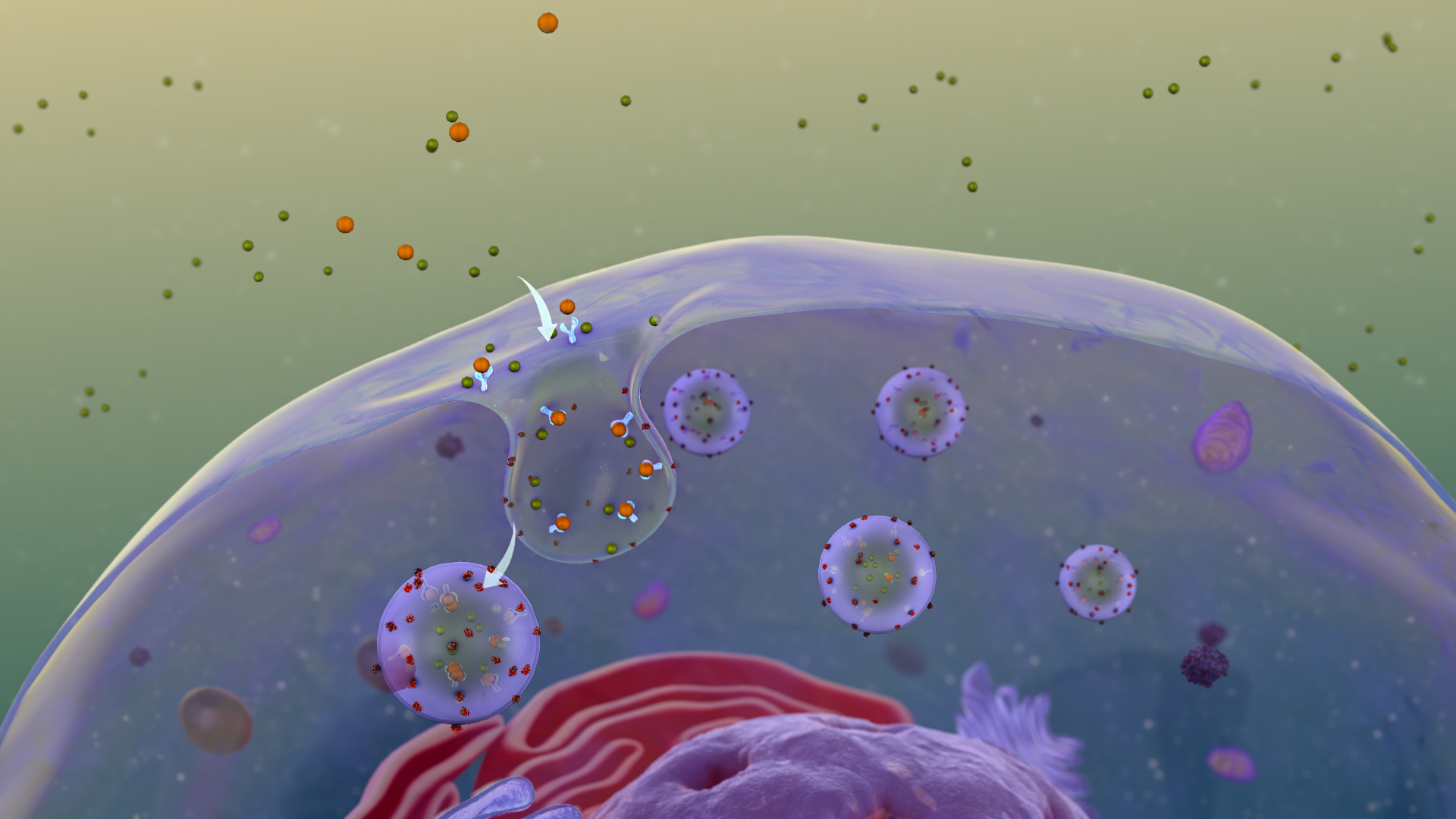
Receptor-mediated endocytosis (RME), also called clathrin-mediated endocytosis, is a process by which cells absorb metabolites,hormones,proteins – and in some cases viruses – by the inward budding of the plasma membrane (invagination). This process forms vesicles containing the absorbed substances and is strictly mediated by receptors on the surface of the cell. Only the receptor-specific substances can enter the cell through this process. (W)
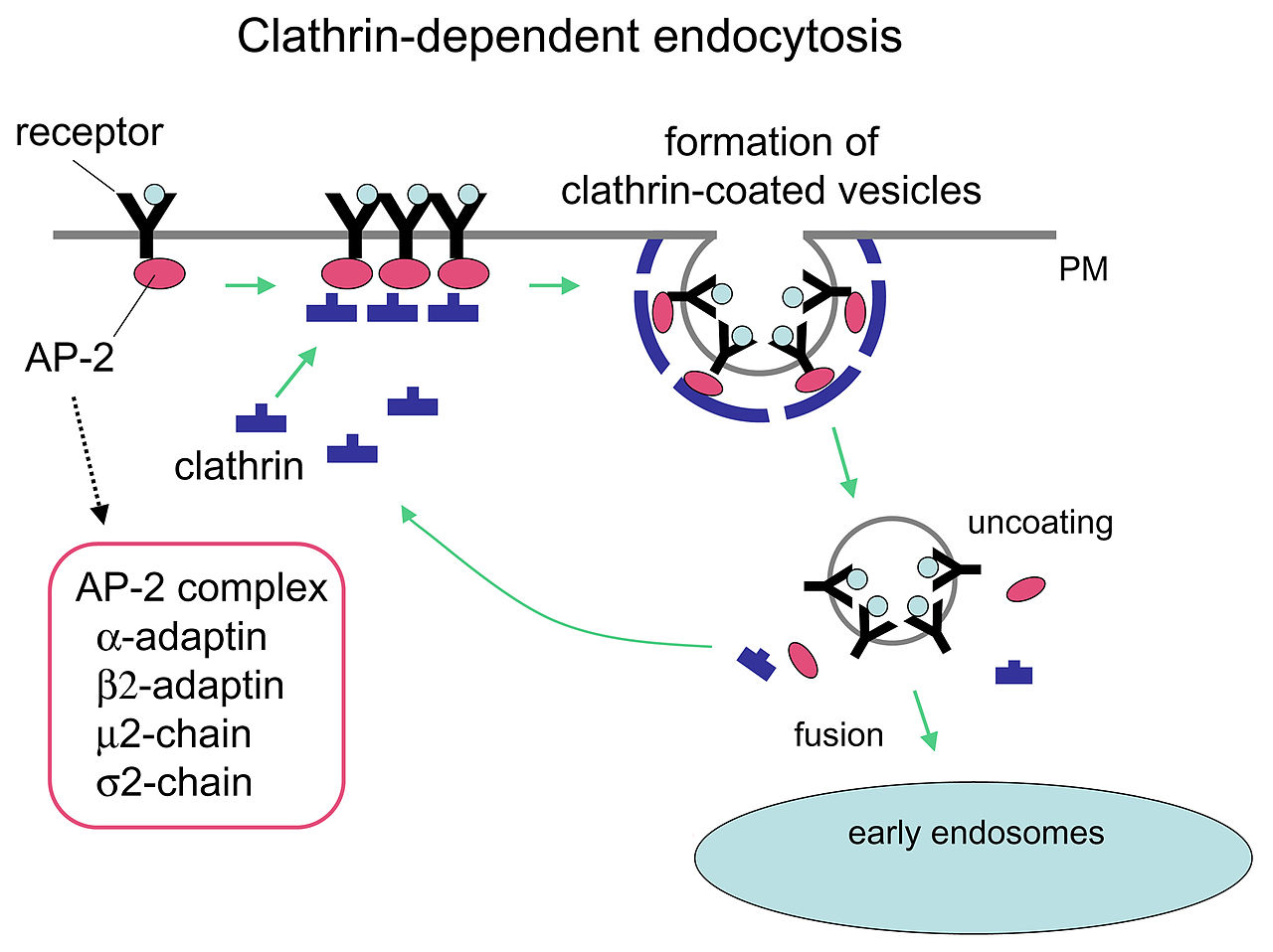
Mechanism of clathrin-dependent endocytosis.
Mechanism of clathrin-dependent endocytosis. Clathrin and cargo molecules are assembled into clathrin-coated pits on the plasma membrane together with an adaptor complex called AP-2 that links clathrin with transmembrane receptors, concluding in the formation of mature clathrin-coated vesicles (CCVs). CCVs are then actively uncoated and transported to early/sorting endosomes.
Endocytosis is triggered when a specific receptor is activated in Receptor-mediated endocytosis.
Receptor Mediated Endocytosis is widely used for the specific uptake of certain substances required by the cell.
The standard definition of a reference range (usually referred to if not otherwise specified) originates in what is most prevalent in a reference group taken from the general (i.e. total) population. This is the general reference range. However, there are also optimal health ranges (ranges that appear to have the optimal health impact) and ranges for particular conditions or statuses (such as pregnancy reference ranges for hormone levels).
Values within the reference range (WRR) are those within the normal distribution and are thus often described as within normal limits (WNL). The limits of the normal distribution are called the upper reference limit (URL) or upper limit of normal (ULN) and the lower reference limit (LRL) or lower limit of normal (LLN). In health care–related publishing, style sheets sometimes prefer the word reference over the word normal to prevent the nontechnical senses of normal from being conflated with the statistical sense. Values outside a reference range are not necessarily pathologic, and they are not necessarily abnormal in any sense other than statistically. Nonetheless, they are indicators of probable pathosis. Sometimes the underlying cause is obvious; in other cases, challenging differential diagnosis is required to determine what is wrong and thus how to treat it.
A cutoff or threshold is a limit used for binary classification, mainly between normal versus pathological (or probably pathological). Establishment methods for cutoffs include using an upper or a lower limit of a reference range. (W)
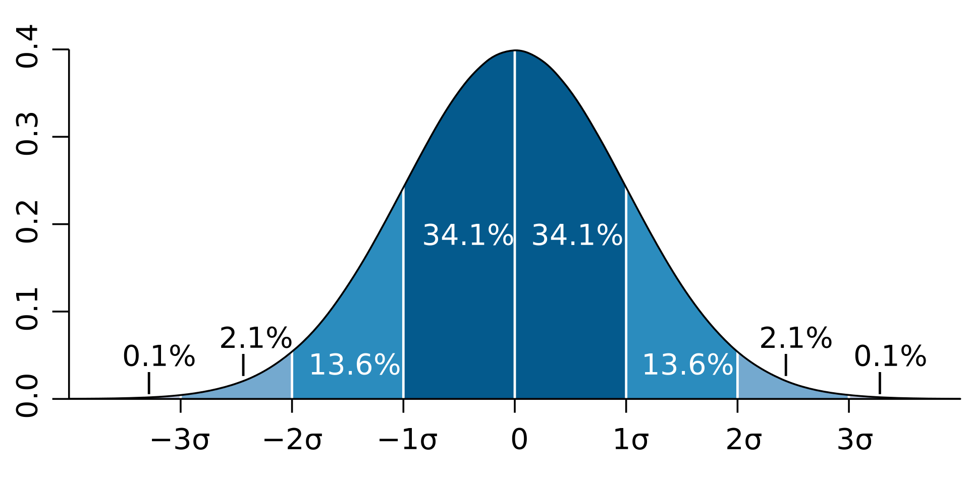
When assuming a normal distribution, the reference range is obtained by measuring the values in a reference group and taking two standard deviations either side of the mean. This encompasses ~95% of the total population.
Normal distribution curve that illustrates standard deviations. Each band has 1 standard deviation, and the labels indicate the approximate proportion of area (note: these add up to 99.8%, and not 100% because of rounding for presentation).
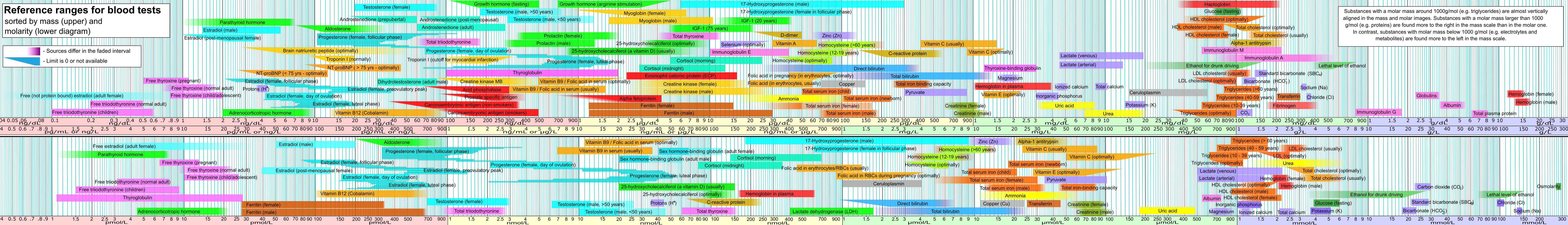
reference ranges for blood tests
Reference ranges for blood tests are sets of values used by a health professional to interpret a set of medical test results from blood samples.Reference ranges for blood tests are studied within the field of clinical chemistry (also known as "clinical biochemistry", "chemical pathology" or "pure blood chemistry"), the area of pathology that is generally concerned with analysis of bodily fluids.
Blood test results should always be interpreted using the reference range provided by the laboratory that performed the test. (W)
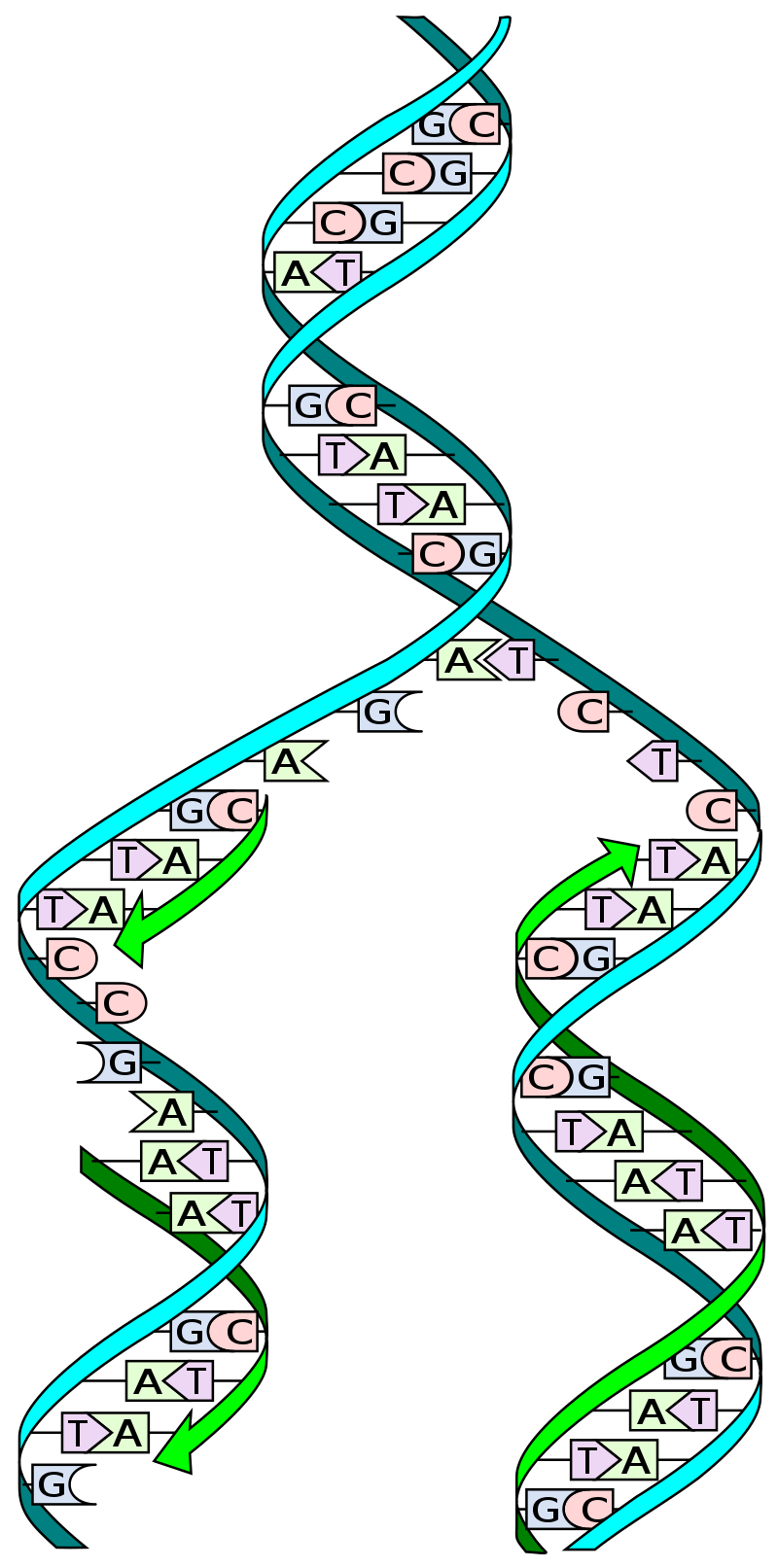
In molecular biology,DNA replication is the biological process of producing two identical replicas of DNA from one original DNA molecule. DNA replication occurs in all living organisms acting as the most essential part for biological inheritance. The cell possesses the distinctive property of division, which makes replication of DNA essential. (W)
DNA replication: The double helix is un'zipped' and unwound, then each separated strand (turquoise) acts as a template for replicating a new partner strand (green). Nucleotides (bases) are matched to synthesize the new partner strands into two new double helices..
Ribosomes assemble polymericproteinmolecules whose sequence is controlled by the sequence of messenger RNA molecules. This is required by all living cells and associated viruses..
ribosome (3D)
The ribosome is a complex composed of RNA and protein that adds up to several million daltons in size and plays a critical role in the process of decoding the genetic information stored in the genome into protein as outlined in what is now known as the Central Dogma of Molecular Biology. Specifically, the ribosome carries out the process of translation, decoding the genetic information encoded in messenger RNA, one amino acid at a time, into newly synthesized polypeptide chains. (W)
Just as higher forms of life have evolved a complex mitotic apparatus to partition duplicated DNA during cell division,bacteria require a specialized apparatus to partition their duplicated DNA. In bacteria, segrosomes perform the function similar to that performed by mitotic spindle. Therefore, segrosomes can be thought of as minimalist spindles.
Segrosomes are usually composed of three basic components- the DNA (plasmid or chromosome) that needs to be segregated into daughter cells, a motor protein that provides the necessary physical forces for accomplishing the segregation and a DNA binding protein that connects the DNA and the motor protein, to form the complete segrosome complex. (W)
senescence
Senescence or biologicalaging is the gradual deterioration of functional characteristics. The word senescence can refer either to cellular senescence or to senescence of the whole organism. Organismal senescence involves an increase in death rates and/or a decrease in fecundity with increasing age, at least in the latter part of an organism's life cycle.
Environmental factors may affect aging, for example, overexposure to ultraviolet radiation accelerates skin aging. Different parts of the body may age at different rates. Two organisms of the same species can also age at different rates, making biological aging and chronological aging distinct concepts. (W)
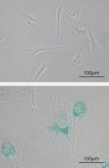
Cellular senescence
(upper) Primary mouse embryonic fibroblast cells (MEFs) before senescence. Spindle-shaped. (lower) MEFs became senescent after passages. Cells grow larger, flatten shape and expressed senescence-associated β-galactosidase (SABG, blue areas), a marker of cellular senescence.
septate junction
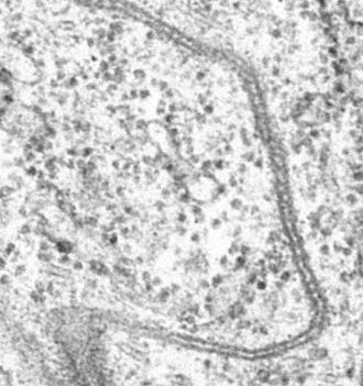
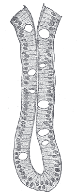
Septate junctions are intercellular junctions found in invertebrate epithelial cells, appearing as ladder-like structures under electron microscopy. They are thought to provide structural strength and a barrier to solute diffusion through the intercellular space. They are considered somewhat analogous to the (vertebrate) tight junctions; however, tight and septate junctions are different in many ways. Known insect homologues of tight junction components are components of conserved signalling pathways that localize to either adherens junctions, the subapical complex, or the marginal zone. Recent studies show that septate junctions are also identified in the myelinated nerve fibers of the vertebrates. (W)
Sepate junction in developing trachea in Drosophyla.
sexual motivation and hormones
Sexual motivation is influenced by hormones such as testosterone,estrogen,progesterone,oxytocin, and vasopressin. In most mammalian species, sex hormones control the ability to engage in sexual behaviors. However, sex hormones do not directly regulate the ability to copulate in primates (including humans). Rather, sex hormones in primates are only one influence on the motivation to engage in sexual behaviours. (W)
skin flora
he term skin flora (also commonly referred to as skin microbiota) refers to the microorganisms which reside on the skin, typically human skin.
Many of them are bacteria of which there are around 1,000 species upon human skin from nineteen phyla. Most are found in the superficial layers of the epidermis and the upper parts of hair follicles.
Skin flora is usually non-pathogenic, and either commensal (are not harmful to their host) or mutualistic (offer a benefit). The benefits bacteria can offer include preventing transient pathogenic organisms from colonizing the skin surface, either by competing for nutrients, secreting chemicals against them, or stimulating the skin's immune system. However, resident microbes can cause skin diseases and enter the blood system, creating life-threatening diseases, particularly in immunosuppressed people.
Depiction of the human body and bacteria that predominate.
sleep
Sleep is a naturally recurring state of mind and body, characterized by altered consciousness, relatively inhibited sensory activity, reduced muscle activity and inhibition of nearly all voluntary muscles during rapid eye movement (REM) sleep, and reduced interactions with surroundings. It is distinguished from wakefulness by a decreased ability to react to stimuli, but more reactive than a coma or disorders of consciousness, with sleep displaying very different and active brain patterns. (W)
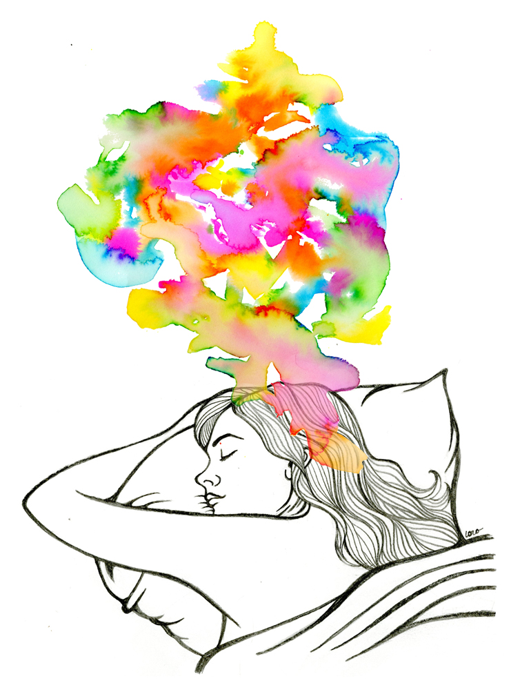
Sleep is associated with a state of muscle relaxation and reduced perception of environmental stimuli. A sleeping girl, 2011.
An artist's creative illustration depicting REM sleep.
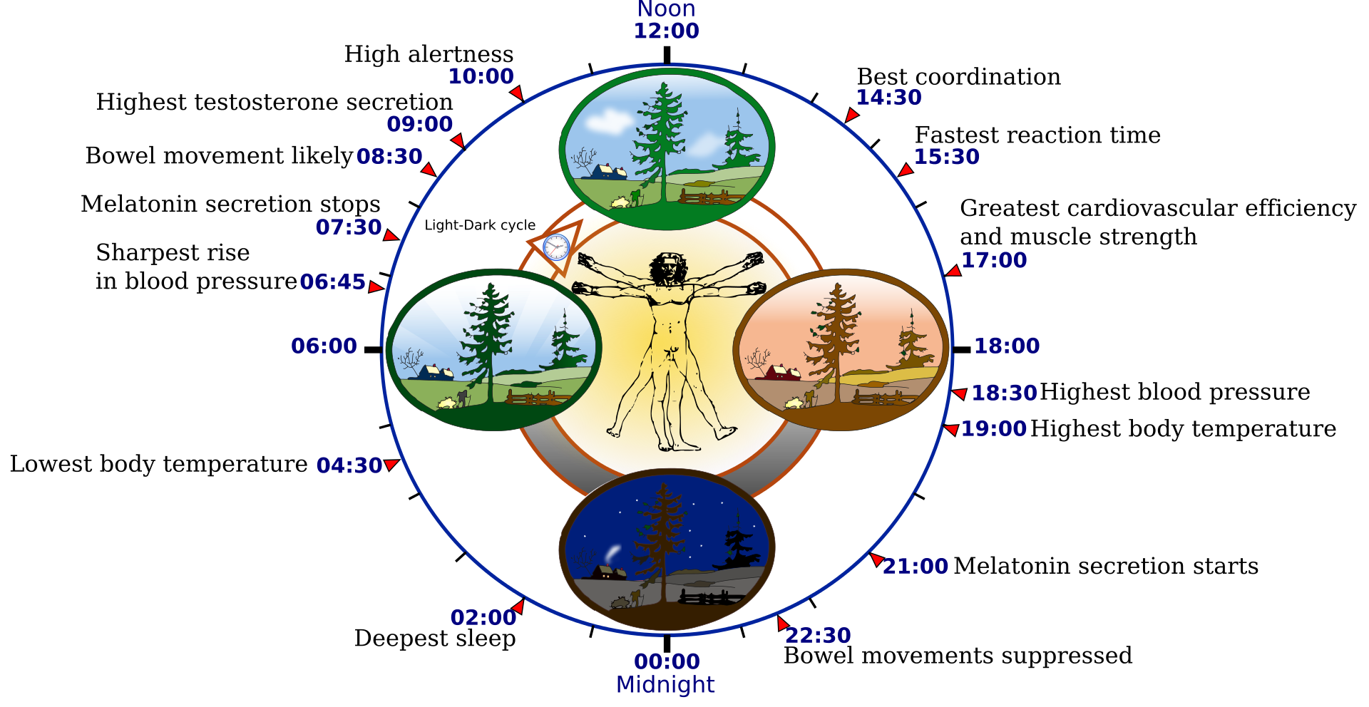
The human "biological clock". Overview of biological circadian clock in humans. Biological clock affects the daily rhythm of many physiological processes. This diagram depicts the circadian patterns typical of someone who rises early in morning, eats lunch around noon, and sleeps at night (10 p.m.). Although circadian rhythms tend to be synchronized with cycles of light and dark, other factors - such as ambient temperature, meal times, stress and exercise - can influence the timing as well. (W)
sorption
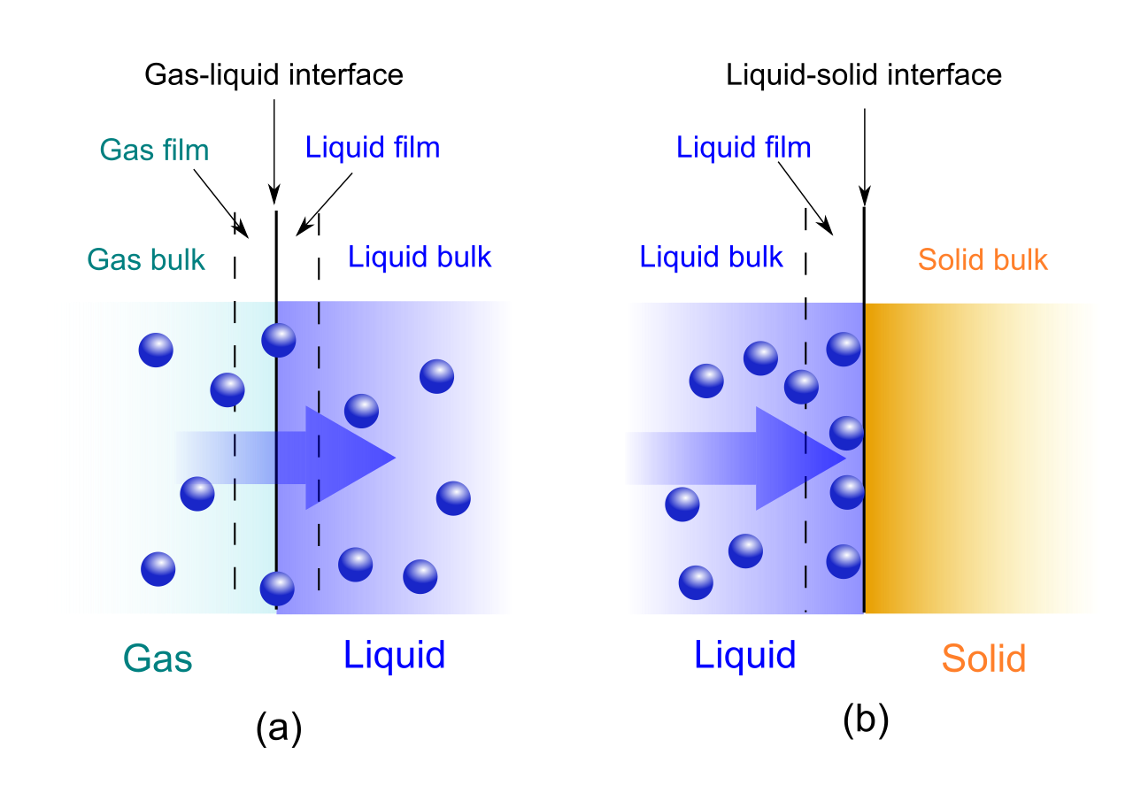
Sorption is a physical and chemical process by which one substance becomes attached to another. Specific cases of sorption are treated in the following articles:
Absorption – "the incorporation of a substance in one state into another of a different state" (e.g., liquids being absorbed by a solid or gases being absorbed by a liquid);
Adsorption – the physical adherence or bonding of ions and molecules onto the surface of another phase (e.g., reagents adsorbed to a solid catalyst surface);
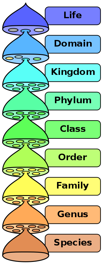
The total number of species is estimated to be between 8 and 8.7 million. However the vast majority of them are not studied or documented and it may take over 1000 years to fully catalogue them all.
Sponges, the members of the phylumPorifera (meaning "pore bearer"), are a basal Metazoa (animal) clade as a sister of the Diploblasts. They are multicellular organisms that have bodies full of pores and channels allowing water to circulate through them, consisting of jelly-like mesohyl sandwiched between two thin layers of cells. The branch of zoology that studies sponges is known as spongiology.(W)
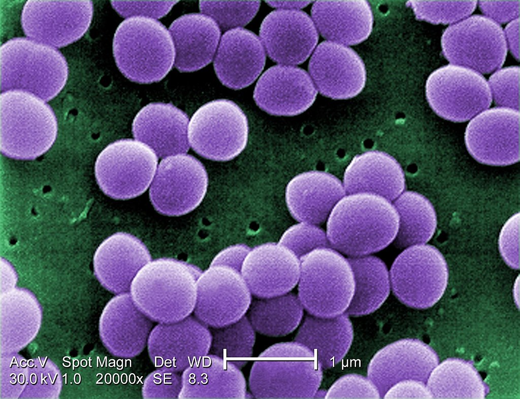
Scanning electron micrograph of "S. aureus"; false color added. Under a very high magnification of 20,000x, this scanning electron micrograph (SEM) shows a strain of Staphylococcus aureus bacteria taken from a vancomycin intermediate resistant culture (VISA). Under SEM, one can not tell the difference between bacteria that are susceptible, or multidrug resistant, but with transmission electron microscopy (TEM), VISA isolates exhibit a thickening in the cell wall that may attribute to their reduced susceptibility to vancomycin . See PHIL 11156 for a black and white version of this image. VISA and VRSA are specific types of antimicrobial-resistant staph bacteria. While most staph bacteria are susceptible to the antimicrobial agent vancomycin some have developed resistance. VISA and VRSA cannot be successfully treated with vancomycin because these organisms are no longer susceptibile to vancomycin. However, to date, all VISA and VRSA isolates have been susceptible to other Food and Drug Administration (FDA) approved drugs. How do VISA and VRSA get their names? Staph bacteria are classified as VISA or VRSA based on laboratory tests. Laboratories perform tests to determine if staph bacteria are resistant to antimicrobial agents that might be used for treatment of infections. For vancomycin and other antimicrobial agents, laboratories determine how much of the agent it requires to inhibit the growth of the organism in a test tube. The result of the test is usually expressed as a minimum inhibitory concentration (MIC) or the minimum amount of antimicrobial agent that inhibits bacterial growth in the test tube. Therefore, staph bacteria are classified as VISA if the MIC for vancomycin is 4-8µg/ml, and classified as VRSA if the vancomycin MIC is >16µg/ml. (W).
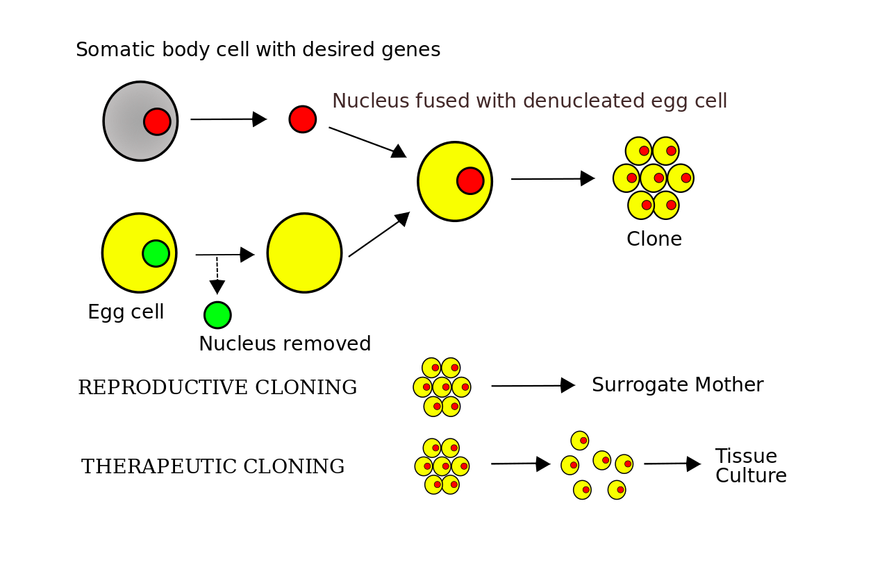
Transplantation of nuclei taken from somatic cells into an oocyte (egg cell) lacking its own nucleus (removed in lab)
Fusion of somatic cells with pluripotent stem cells and
Transformation of somatic cells into stem cells, using the genetic material encoding reprogramming protein factors, recombinant proteins; microRNA, a synthetic, self-replicating polycistronic RNA and low-molecular weight biologically active substances. (W)
Induced totipotent cells usually can be obtained by reprogramming somatic cells by somatic-cell nuclear transfer (SCNT).
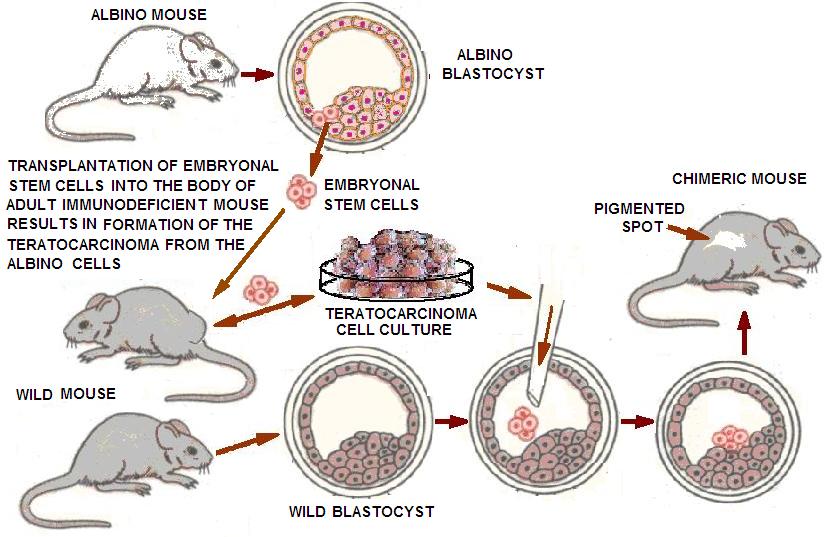
Transplantation of pluripotent/embryonic stem cells into the body of adult mammals, usually leads to the formation of teratomas, which can then turn into a malignant tumor teratocarcinoma. However, putting teratocarcinoma cells into the embryo at the blastocyst stage, caused them to become incorporated in the cell mass and often produced a normal healthy chimeric (i.e. composed of cells from different organisms) animal.
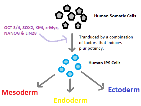
Human somatic cells are made pluripotent by transducing them with factors that induces pluripotency (OCT 3/4, SOX2, Klf4, c-Myc, NANOG and LIN28). This results in the production of IPS cells, which can differentiate into any cells of the three embryonic germ layers (Mesoderm, Endoderm, Ectoderm).
An intestinal crypt - an accessible and abundant source of intestinal epithelial cells for conversion into β-like cells.
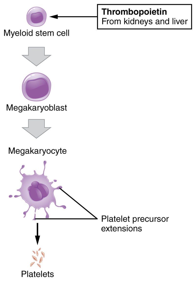
Platelets extruded from megakaryocytes.
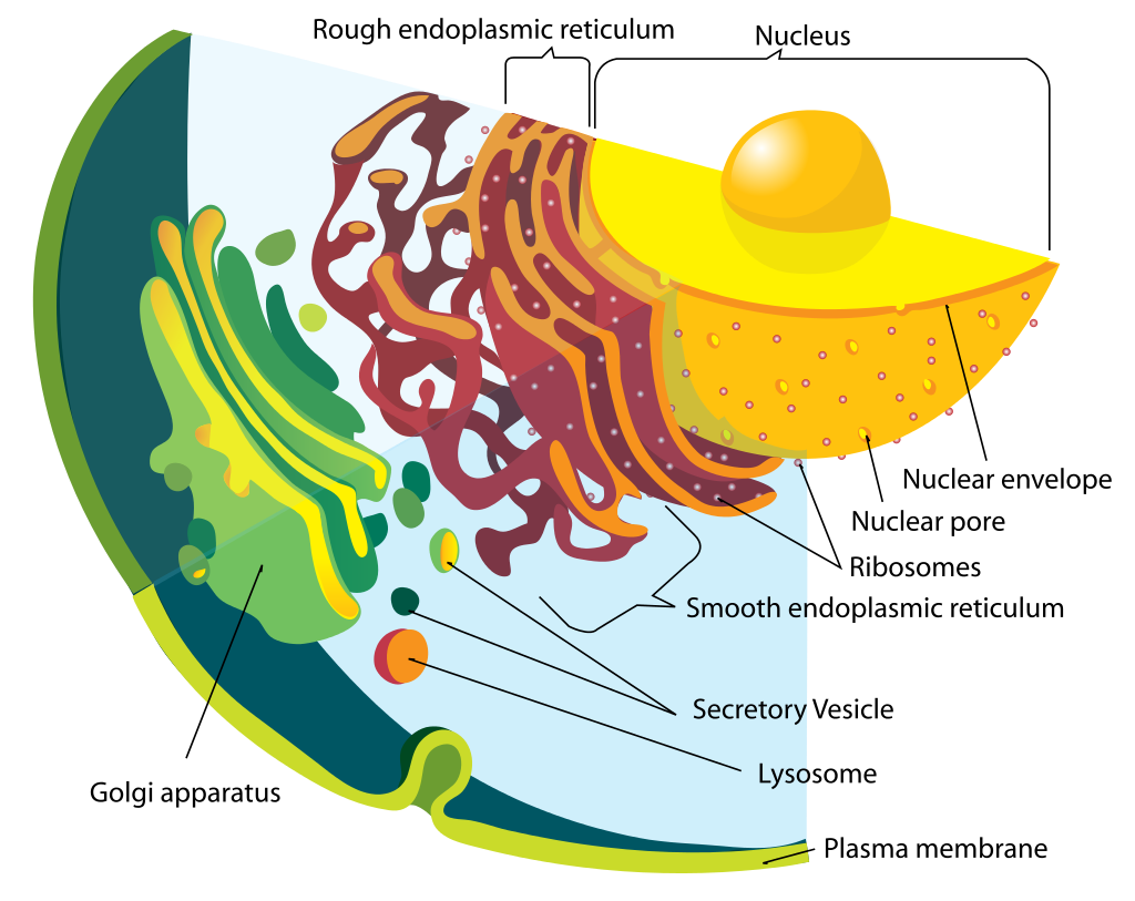
Illustration from Anatomy & Physiology, Connexions Web site. http://cnx.org/content/col11496/1.6/, Jun 19, 2013.
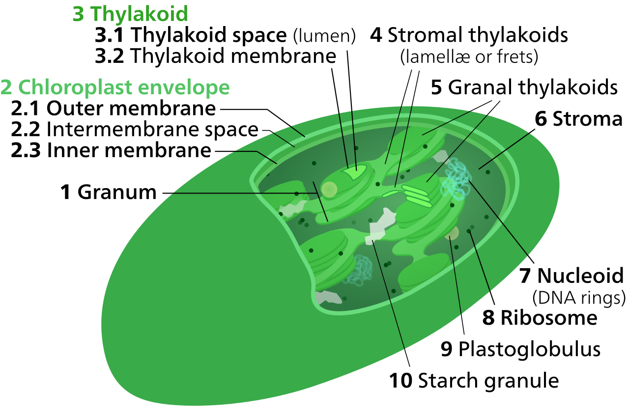
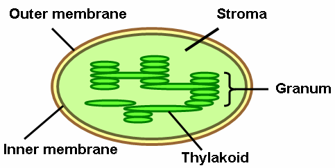
Within the stroma are grana (stacks of thylakoid), and the sub-organelles or daughter cells, where photosynthesis is commenced before the chemical changes are completed in the stroma.
Chloroplast diagram designed for small sizes with text and number labels. Isn't it so cute?
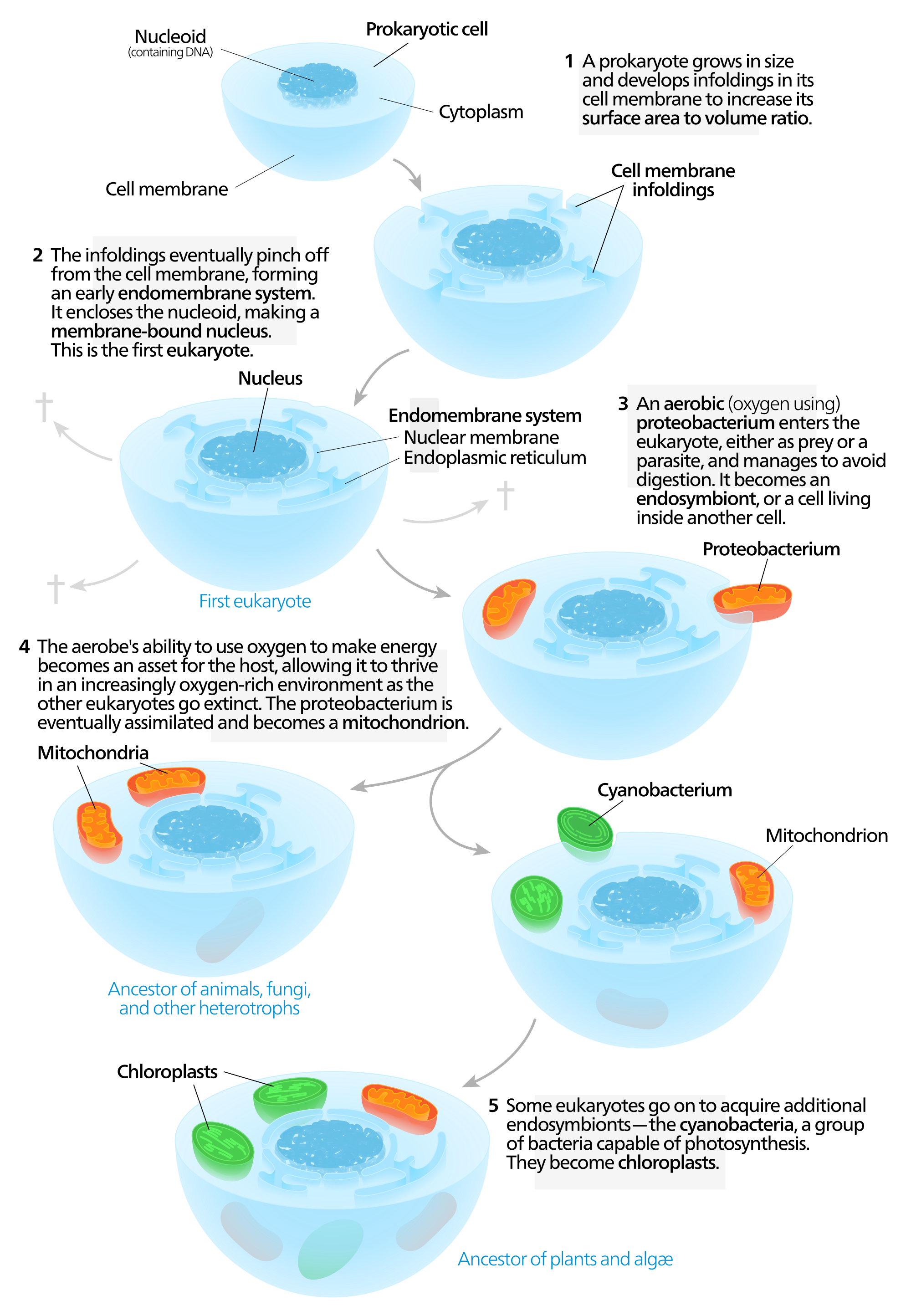
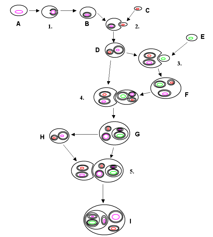
symbiogenesis Symbiogenesis, or endosymbiotic theory, is an evolutionary theory of the origin of eukaryotic cells from prokaryotic organisms, first articulated in 1905 and 1910 by the Russian botanist Konstantin Mereschkowski, and advanced and substantiated with microbiological evidence by Lynn Margulis in 1967. It holds that the organelles distinguishing eukaryote cells evolved through symbiosis of individual single-celled prokaryotes (bacteria and archaea). The theory holds that mitochondria,plastids such as chloroplasts, and possibly other organelles of eukaryotic cells are descended from formerly free-living prokaryotes taken one inside the other in endosymbiosis. Mitochondria appear to be phylogenetically related to Rickettsialesproteobacteria, and chloroplasts to nitrogen-fixing filamentous cyanobacteria. Among the many lines of evidence supporting symbiogenesis are that new mitochondria and plastids are formed only through binary fission, and that cells cannot create new ones otherwise; that the transport proteins called porins are found in the outer membranes of mitochondria, chloroplasts and bacterial cell membranes; that cardiolipin is found only in the inner mitochondrial membrane and bacterial cell membranes; and that some mitochondria and plastids contain single circular DNA molecules similar to the chromosomes of bacteria. (W)
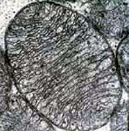
Electron micrograph of a single mitochondrion showing the organized arrangement of the protein matrix and the inner mitochondrial membranes. (Photo: U.S. Dept. of Health and Human Services/National Institutes of Health).
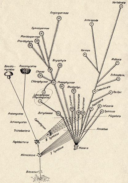
Symbiogenesis tree of life diagram by Konstantin Mereschkowski (1855–1921), showing the origin of complex life-forms by two episodes of incorporation of symbioticbacteria, 1905. The first symbiosis generated the nucleus. The second symbiosis generated chloroplasts. Mitochondria were not included.
One model for the origin of mitochondria and plastids.
One of many models on where eukaryotes came from. This model has an amitochondriate eukaryote engulfing an aerobe and then a cyanobacterium. Includes a build-your own SVG eukaryote kit with assorted mitochondria and plastids. (off the page) :P —Sources— 1 Biology, 8th edition Campbell & Reece—page 517 2, 3.
Modern endosymbiotic theory posits that simple life forms merged, forming cell organelles, like mitochondria.
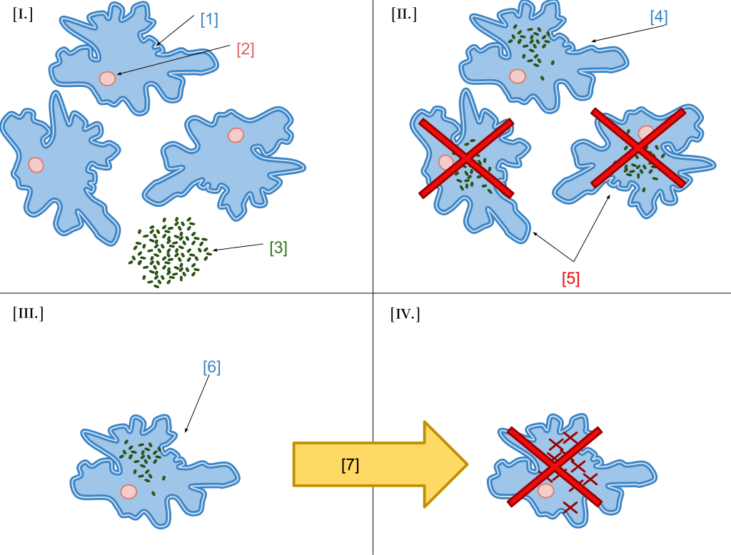
Kwang Jeon's experiment: [I] Amoebae infected by x-bacteria [II] Many amoebae become sick and die [III] Survivors have x-bacteria living in their cytoplasm [IV] Antibiotics kill x-bacteria: host amoebae die as now dependent on x-bacteria.
In 1966 Microbiologist Kwang Jeon conducted an experiment with amoebae communities providing real-life evidence for the endosymbiotic theory. The single-celled amoebae community was infected by a bacterial infection of x-bacteria. The x-bacteria caused many of the amoebae to become sick and die. Very few of the amoebae survived the epidemic. Amoebae surviving the epidemic have x-bacteria living inside their cytoplasm. Antibiotics were used to kill the x-bacteria inside the remaining amoebae causing the host amoeba to die as well because the amoebae had become dependant on the x-bacteria to survive. Labels: [1] Amoebae, [2] Nucleos, [3] X-Bacteria, [4] Infected Amoebae, [5] Sick Amoebae, [6] Surviving Amoebae, [7] Add Antibiotic.
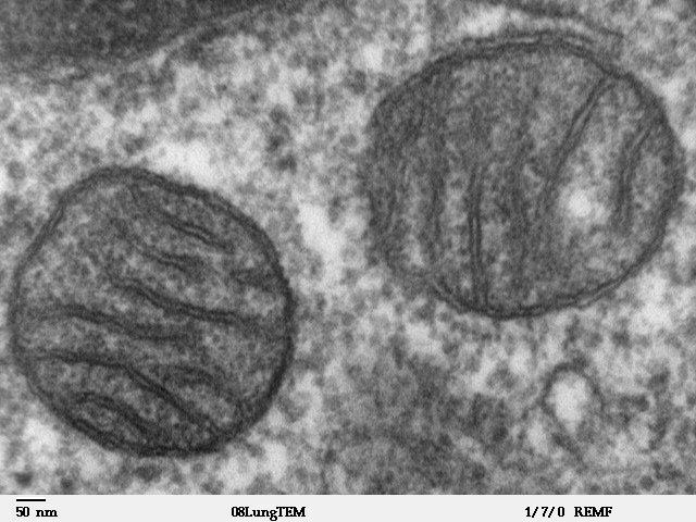
Mitochondria of a mammal lung cell visualized using Transmission Electron Microscopy.
Transmission electron microscope image of a thin section cut through an area of mammalian lung tissue. The high magnification image shows a mitochondria. JEOL 100CX TEM.
Diagram of endomembrane system in eukaryotic cell.
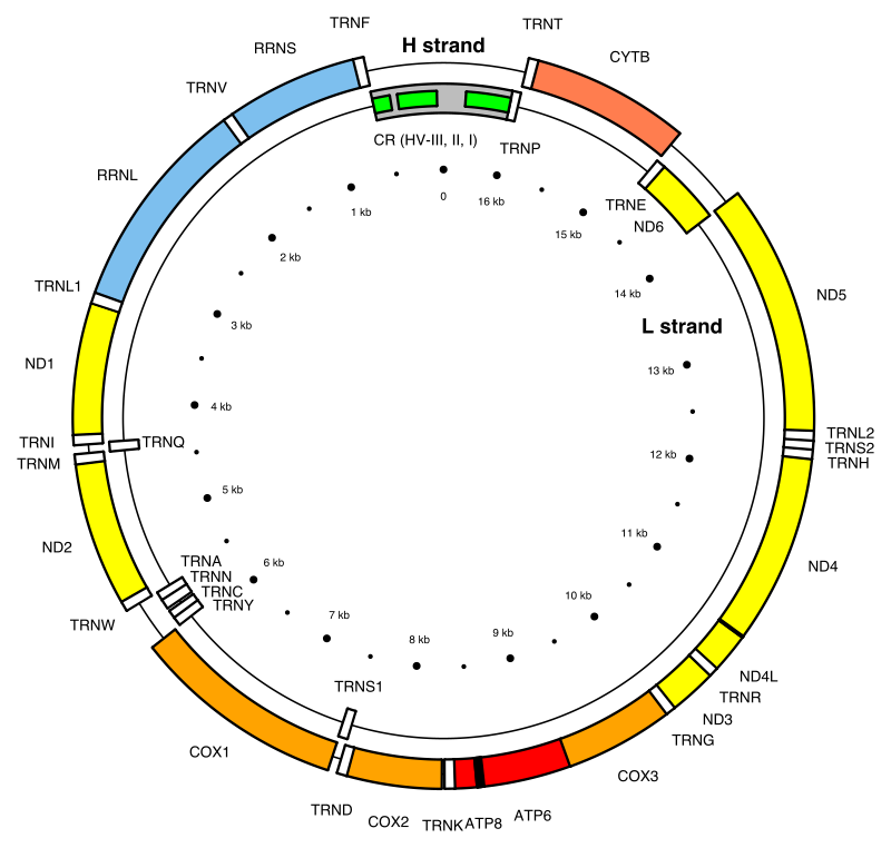
Map of the human mitochondrial DNA genome (16569 bp, NCBI sequence accession NC_012920 — Anderson et al. 1981). The H (heavy, outer circle) and L (light, inner circle) strands are given with their corresponding genes. There are 22 transfer RNA (TRN) genes for the following amino acids: F, V, L1 (codon UUA/G), I, Q, M, W, A, N, C, Y, S1 (UCN), D, K, G, R, H, S2 (AGC/U), L2 (CUN), E, T and P (white boxes). There are 2 ribosomal RNA (RRN) genes: S (small subunit, or 12S) and L (large subunit, or 16S) (blue boxes). There are 13 protein-coding genes: 7 for NADH dehydrogenase subunits (ND, yellow boxes), 3 for cytochrome c oxidase subunits (COX, orange boxes), 2 for ATPase subunits (ATP, red boxes), and one for cytochrome b (CYTB, coral box). Two gene overlaps are indicated (ATP8-ATP6, and ND4L-ND4, black boxes). The control region (CR) is the longest non-coding sequence (grey box). Its three hyper-variable regions are indicated (HV, green boxes)..
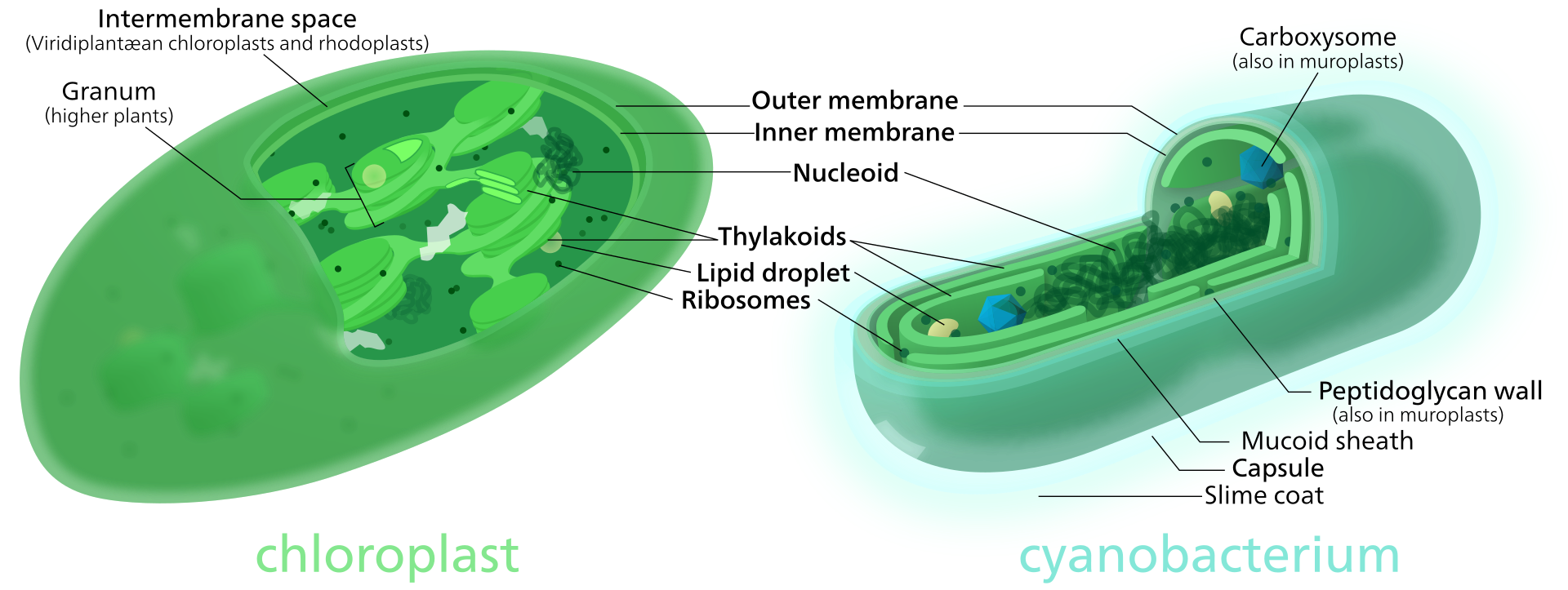
Comparison between a chloroplast and a cyanobacterium. Lines aligned to a 690 × 260 px grid, Librsvg bugs begin below about 515 × 195 px.
synapse
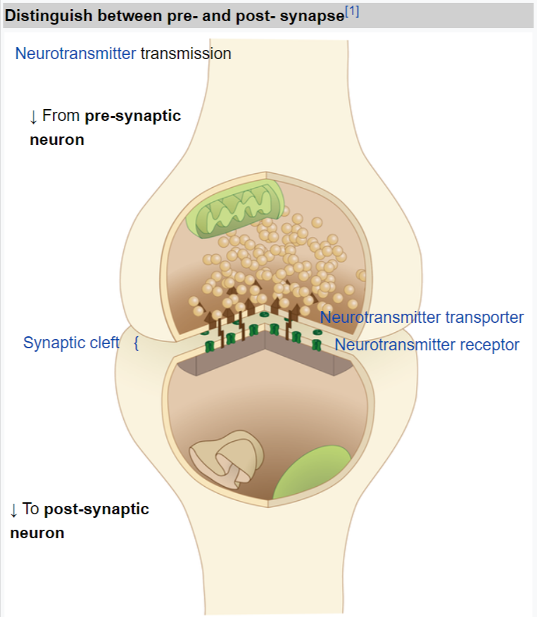
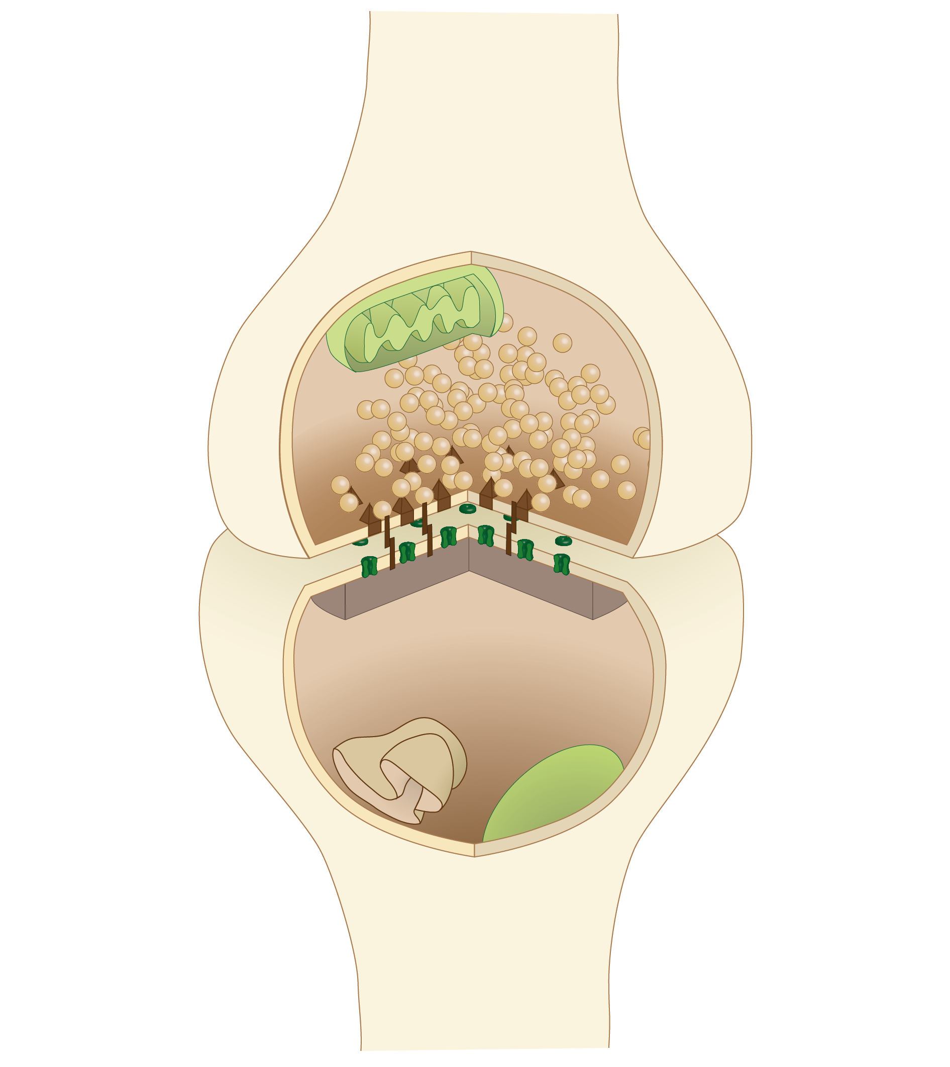
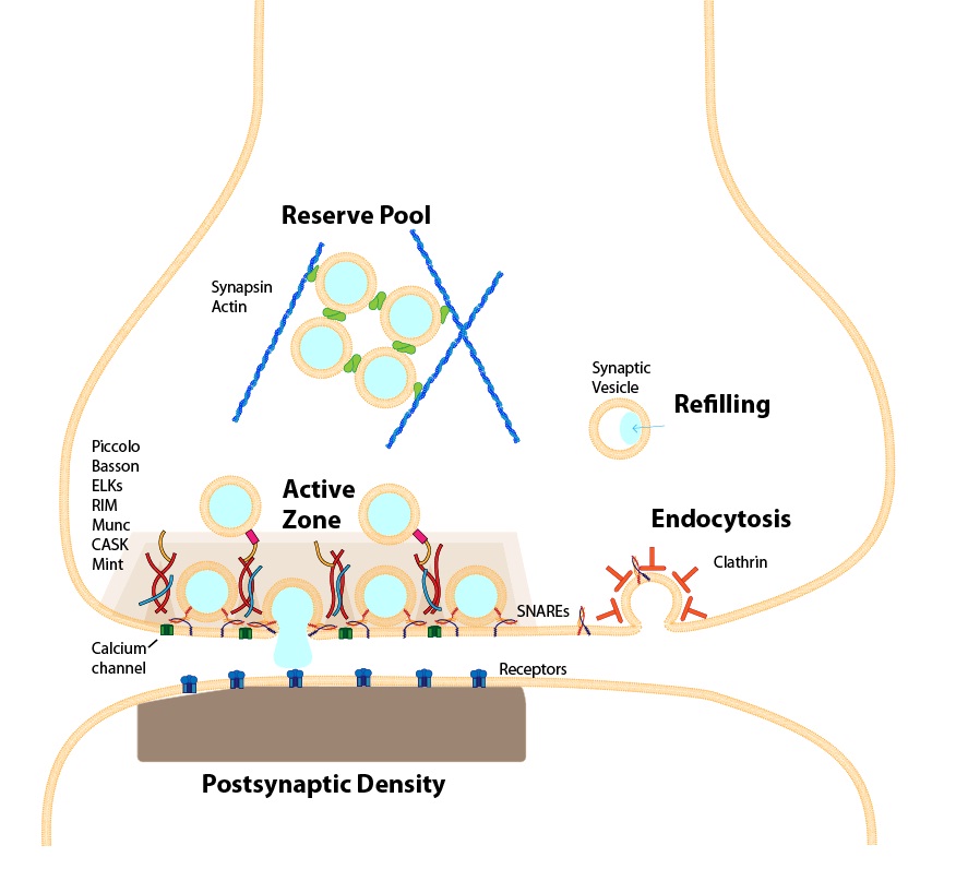
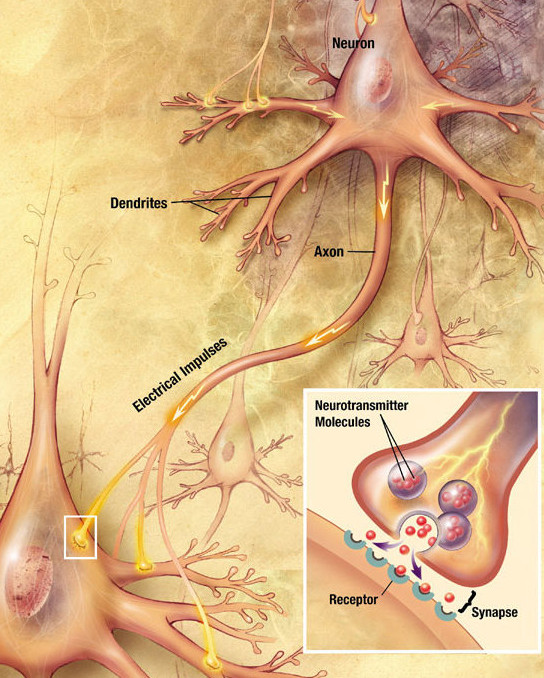
In the nervous system, a synapse is a structure that permits a neuron (or nerve cell) to pass an electrical or chemical signal to another neuron or to the target effector cell.
Santiago Ramón y Cajal proposed that neurons are not continuous throughout the body, yet still communicate with each other, an idea known as the neuron doctrine. The word "synapse" – from the Greeksynapsis (συνάψις), meaning "conjunction", in turn from συνάπτεὶν (συν ("together") and ἅπτειν ("to fasten")) – was introduced in 1897 by the English neurophysiologist Charles Sherrington in Michael Foster's Textbook of Physiology. Sherrington struggled to find a good term that emphasized a union between two separate elements, and the actual term "synapse" was suggested by the English classical scholar Arthur Woollgar Verrall, a friend of Foster. Some authors generalize the concept of the synapse to include the communication from a neuron to any other cell type, such as to a motor cell, although such non-neuronal contacts may be referred to as junctions (a historically older term). A landmark study by Sanford Palay demonstrated the existence of synapses.
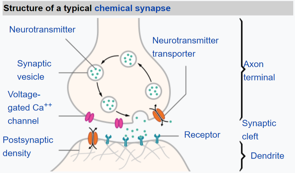
Synapses are essential to neuronal function: neurons are cells that are specialized to pass signals to individual target cells, and synapses are the means by which they do so. At a synapse, the plasma membrane of the signal-passing neuron (the presynaptic neuron) comes into close apposition with the membrane of the target (postsynaptic) cell. Both the presynaptic and postsynaptic sites contain extensive arrays of molecular machinery that link the two membranes together and carry out the signaling process. In many synapses, the presynaptic part is located on an axon and the postsynaptic part is located on a dendrite or soma.Astrocytes also exchange information with the synaptic neurons, responding to synaptic activity and, in turn, regulating neurotransmission. Synapses (at least chemical synapses) are stabilized in position by synaptic adhesion molecules (SAMs) projecting from both the pre- and post-synaptic neuron and sticking together where they overlap; SAMs may also assist in the generation and functioning of synapses. (W)
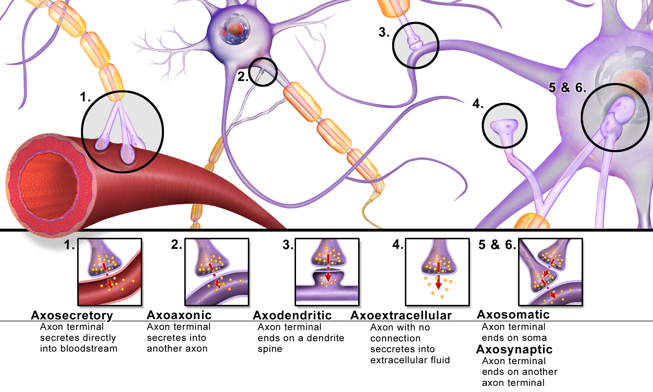
"The connection linking neuron to neuron is the synapse. Signal flows
in one direction, from the presynaptic neuron to the postsynaptic neuron
via the synapse which acts as a variable attenuator." In brief,
the direction of the signal flow determines the prefix for the involved
synapses..
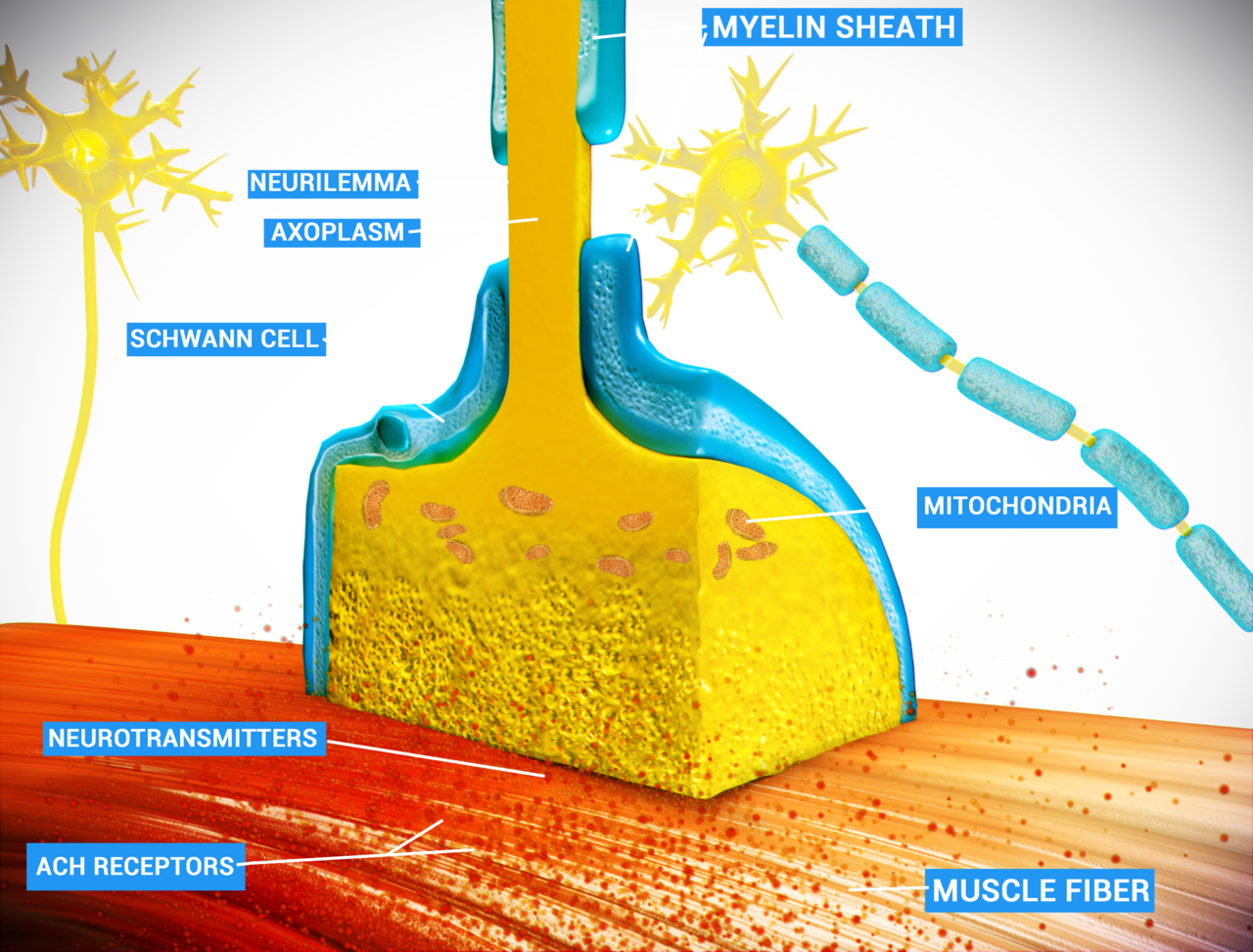
A neuromuscular junction (or myoneural junction) is a chemical synapse formed by the contact between a motor neuron and a muscle fiber. It is at the neuromuscular junction that a motor neuron is able to transmit a signal to the muscle fiber, causing muscle contraction.
📹 Chemical Synapse — Cholinergic Synapse / blausen (LINK)
📌 TRANSCRIPTION
A neuron relays information to another neuron at a synapse. The neuron transmitting the information is called the pre-synaptic neuron, and the neuron receiving the information is the postsynaptic neuron. Let’s look at what happens at a cholinergic synapse, one of the most common types of synapses. When an action potential arrives at the synaptic knob of the pre-synaptic neuron, voltage-regulated calcium gates open, calcium ions enter and bind to synaptic vesicles. This leads to exocytosis which releases the neurotransmitter acetylcholine from the synaptic vesicles into the synaptic cleft. Acetylcholine molecules then diffuse across the synaptic cleft and bind to ACh receptors in the membrane of the postsynaptic neuron. The binding opens ion channels, and the membrane depolarizies. If this depolariazation brings the initial segment of the postsynaptic neuron to threshold, it will result in an action potential. The depolarization of the postsynaptic membrane is short-lived because acetylcholinesterase rapidly breaks down the ACh in the synaptic cleft.
📹 Chemical Synapse — Cholinergic Synapse at Neuromuscular Junction / blausen (LINK)
📌 TRANSCRIPTION
A cholinergic synapse is specialized cell-to-cell junction where nerve impulses are transmitted via the release of the neurotransmitter, acetylcholine, from one neuron to another neuron or to a non-neuronal cell, such as a skeletal muscle cell.
t
T cell
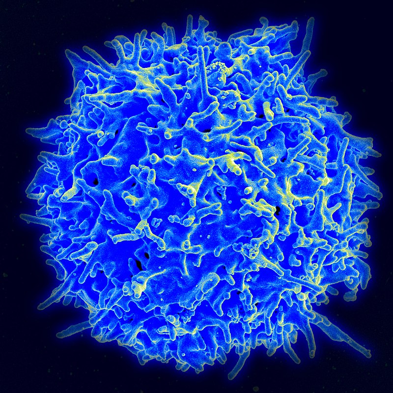
A T cell is a type of lymphocyte, which develops in the thymus gland (hence the name) and plays a central role in the immune response. T cells can be distinguished from other lymphocytes by the presence of a T-cell receptor on the cell surface. These immune cells originate as precursor cells, derived from bone marrow, and develop into several distinct types of T cells once they have migrated to the thymus gland. T cell differentiation continues even after they have left the thymus. (W)
Scanning electron micrograph of a human T lymphocyte (also called a T cell) from the immune system of a healthy donor.
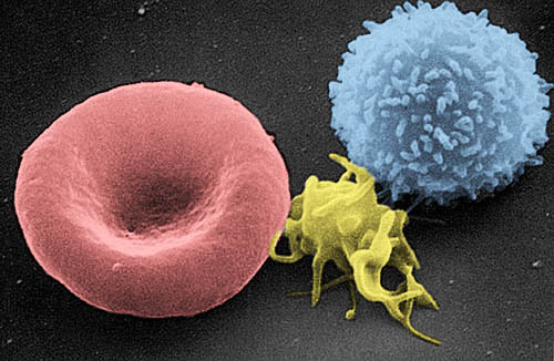
Scanning electron micrograph of a red blood cell (left), a platelet (center), and a T lymphocyte (right).
3D rendering of a T cell.
3D illustration of a lymphocye cell
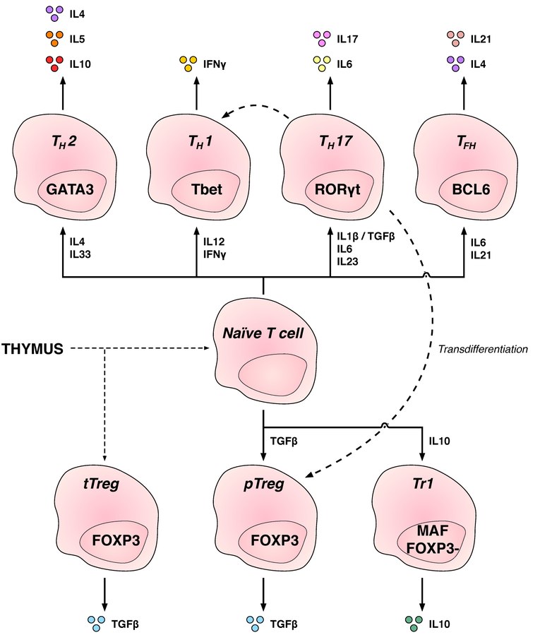
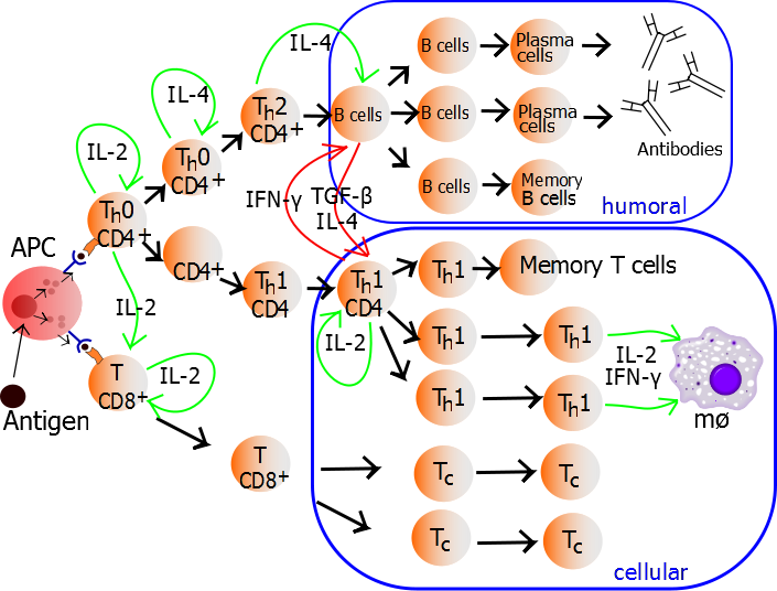
Depiction of the various key subsets of CD4-positive T cells with corresponding associated cytokines and transcription factors.
T cells leave the thymus in a naïve, antigen non-experiened state. After contact with antigen, T cells may take on one of numerous subsets. These subsets are defined by their cytokine secretion profiles and the transcription factors necessary for their development. Dashed line shows reported transdifferentiation between subsets. Effector subsets are shown at the top of the diagram whilst regulatory subsets are shown at the bottom. (W).
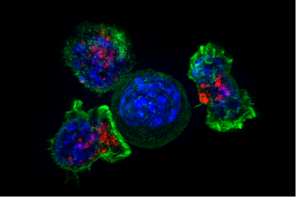
Superresolution image of a group of cytotoxic T cells surrounding a cancer cell.
Superresolution image of a group of killer T cells (green and red) surrounding a cancer cell (blue, center). When a killer T cell makes contact with a target cell, the killer cell attaches and spreads over the dangerous target. The killer cell then uses special chemicals housed in vesicles (red) to deliver the killing blow. This event has thus been nicknamed “the kiss of death”. After the target cell is killed, the killer T cells move on to find the next victim. .
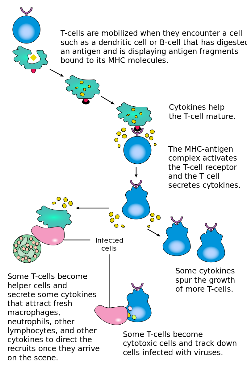
The T lymphocyte activation pathway: T cells contribute to immune defenses in two major ways; some direct and regulate immune responses; others directly attack infected or cancerous cells.
The T lymphocyte activation pathway is triggered when a T cell encounters its cognate antigen, coupled to a MHC molecule, on the surface of an infected cell or a phagocyte. T cells contribute to immune defenses in two major ways: some direct and regulate immune responses; others directly attack infected or cancerous cells.
T helper cell
The T helper cells (Th cells), also known as CD4+ cells, are a type of T cell that play an important role in the immune system, particularly in the adaptive immune system. As their name suggests, they "help" the activity of other immune cells by releasing cytokines, small protein mediators that alter the behavior of target cells that express receptors for those cytokines. These cells help to polarize the immune response into the appropriate kind depending on the nature of the immunological insult (virus vs. extracellular bacterium vs. intracellular bacterium vs. helminth vs. fungus vs. protist). They are generally considered essential in B cellantibody class switching, breaking cross-tolerance in dendritic cells, in the activation and growth of cytotoxic T cells, and in maximizing bactericidal activity of phagocytes such as macrophages and neutrophils.
Mature Th cells express the surface protein CD4 and are referred to as CD4+ T cells. Such CD4+ T cells are generally treated as having a pre-defined role as helper T cells within the immune system. For example, when an antigen-presenting cell displays a peptide antigen on MHC class II proteins, a CD4+ cell will aid those cells through a combination of cell to cell interactions (e.g. CD40 (protein) and CD40L) and through cytokines.(W)
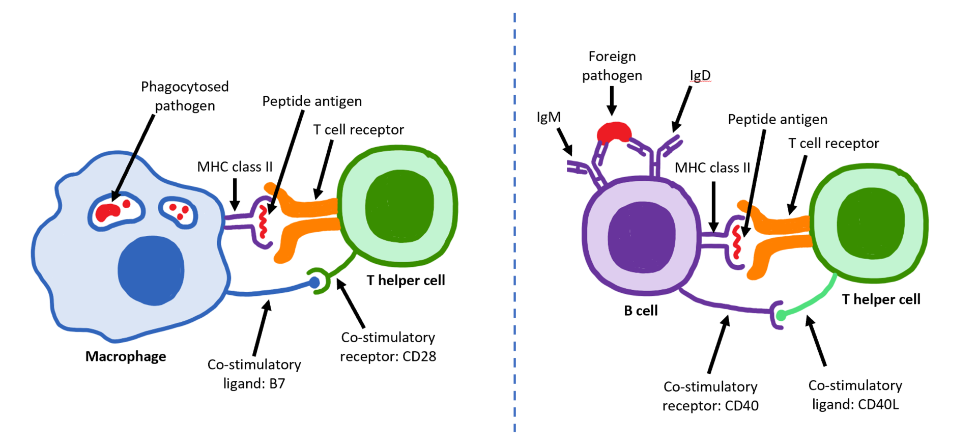
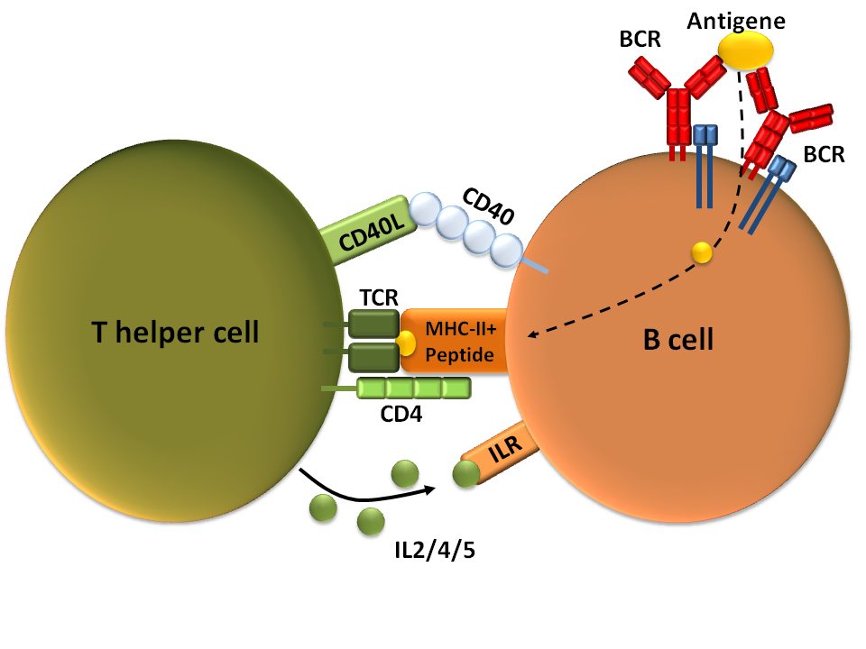
Activation of B cell by T helper cell
This is a visual depicting how T helper cells and B cells are activated. For T cell activation, there must be binding of the T cell receptor to both the antigen peptide and the MHC class II molecule on an antigen presenting cell (APC). Additionally, there must be binding of the two co-stimulatory molecules (B7 on the APC and CD28 on the T cell). For B cell activation, a pathogen must bind to the IgM and IgD antibodies in order to be internalized and presented on the MHC class II molecule of the B cell. Like T cell activation, there must be binding of the two co-stimulatory molecules (in this case CD40 with CD40L). Once a B cell is activated, it turns into a plasma cell which secretes antibodies.
T-cell dependent b-cell activation, showing TH2-cell (left) B-cell (right) and several interaction molecules self-made according to Janeway et al, Immunologie (Berlin, 2002).
T-cell dependent b-cell activation, showing TH2-cell (left) B-cell (right) and several interaction molecules self-made according to: Charles A. Janeway jr. u. a.: Immunologie. 5. Auflage. Spektrum Akademischer Verlag Gmbh, Heidelberg, Berlin 2002, ISBN 3-8274-1078-9.
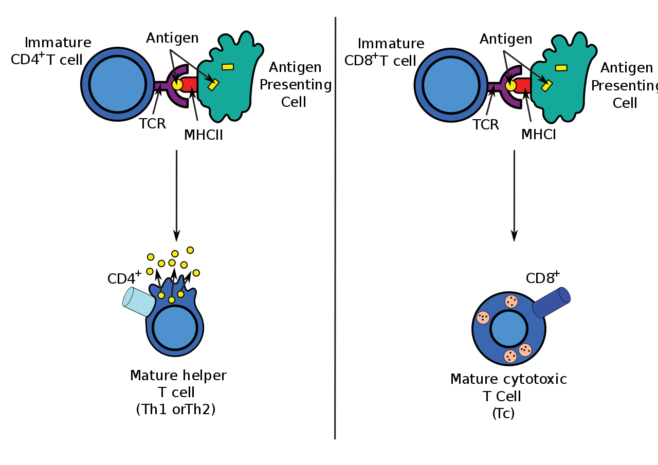
Antigen presentation stimulates naïve CD8+ and CD4+ T cells to respectively become mature "cytotoxic" CD8+ cells and "helper" CD4+ cells..
Antigen presentation stimulates T cells to activate "cytotoxic" CD8+ cells or "helper" CD4+ cells. Cytotoxic cells directly attack other cells carrying certain foreign or abnormal molecules on their surfaces. Helper T cells, or Th cells, coordinate immune responses by communicating with other cells. In most cases, T cells only recognize an antigen if it is carried on the surface of a cell by one of the body’s own MHC, or major histocompatibility complex, molecules.
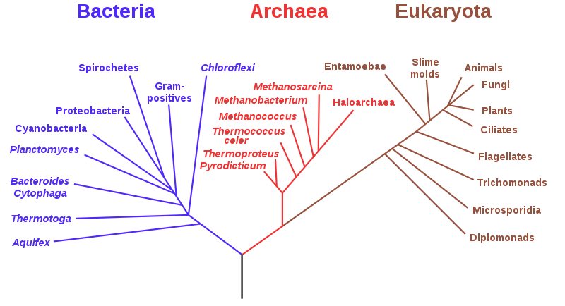
A phylogenetic tree based on rRNA data, emphasizing the separation of bacteria, archaea, and eukaryotes, as proposed by Carl Woese et al. in 1990.
thylakoid
Thylakoids are membrane-bound compartments inside chloroplasts and cyanobacteria. They are the site of the light-dependent reactions of photosynthesis. Thylakoids consist of a thylakoid membrane surrounding a thylakoid lumen. Chloroplast thylakoids frequently form stacks of disks referred to as grana (singular: granum). Grana are connected by intergranal/ stroma thylakoids, which join granum stacks together as a single functional compartment. (W)
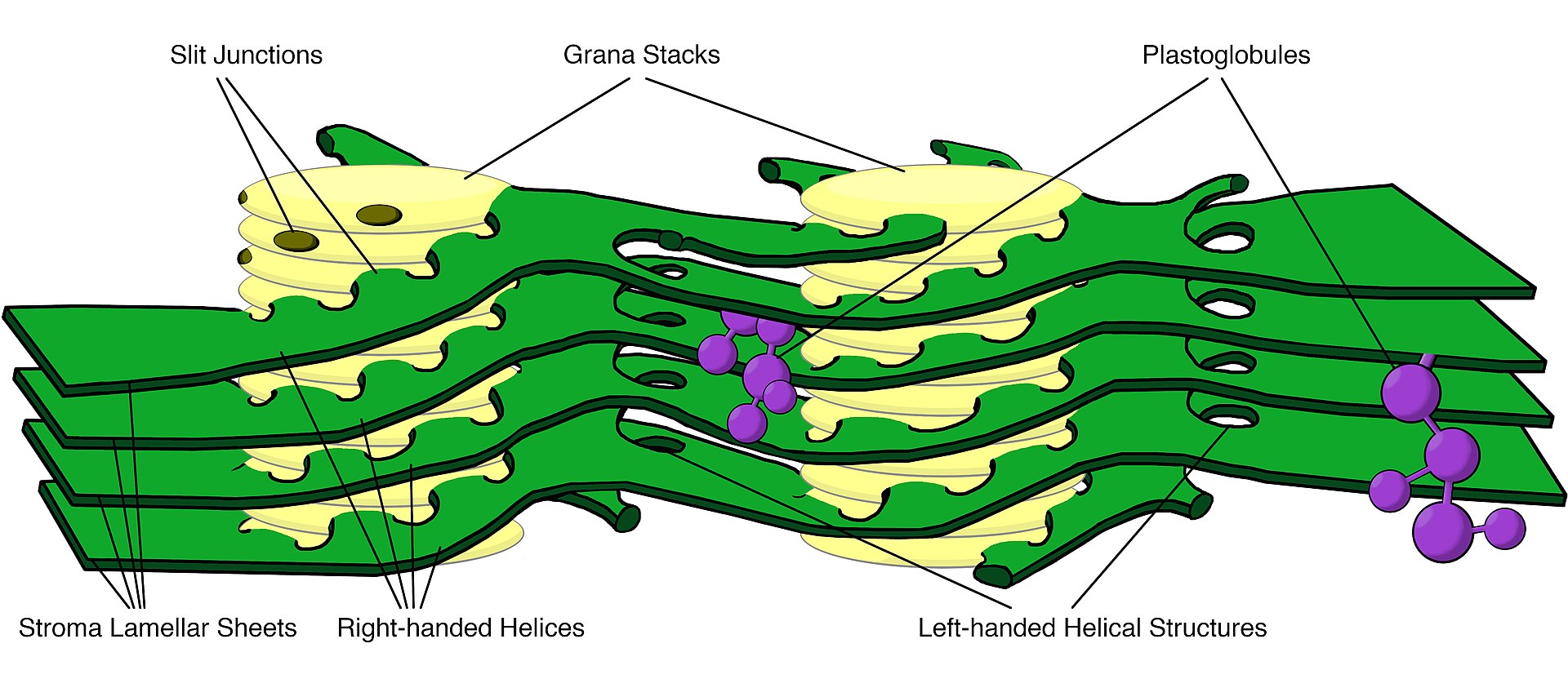
Granum-stroma assembly structure The prevailing model of the granum-stroma assembly is stacks of granal thylakoids wrapped by right-handed helical stromal thylakoids which are connected to large parallel sheets of stromal thylakoids and adjacent right-handed helices by left-handed helical structures.
tight junction
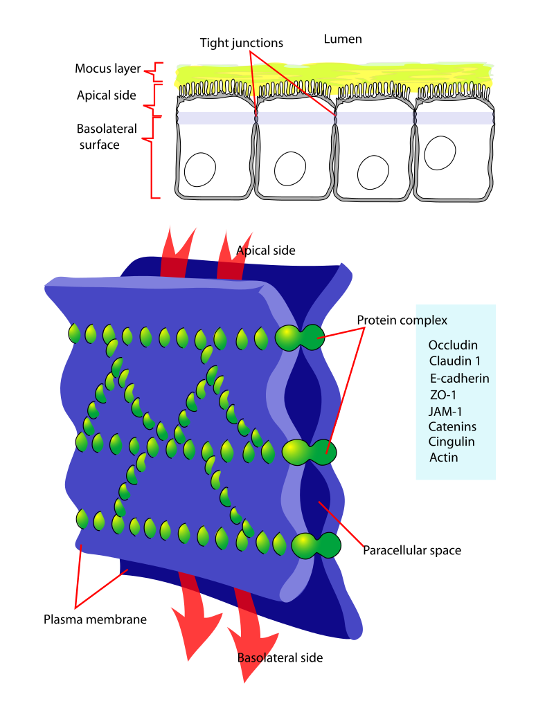
Tight junctions, also known as occluding junctions or zonulae occludentes (singular, zonula occludens) are multiprotein junctional complexes whose general function is to prevent leakage of transported solutes and water and seals the paracellular pathway. Tight junctions may also serve as leaky pathways by forming selective channels for small cations, anions, or water. Tight junctions are present mostly in vertebrates (with the exception of Tunicates). The corresponding junctions that occur in invertebrates are septate junctions.(W)
Diagram showing a tight junction. Tight junctions seal adjacent epithelial cells in a narrow band just beneath their apical surface.
Depiction of the transmembrane proteins that make up tight junctions: occludin, claudins, and JAM proteins.
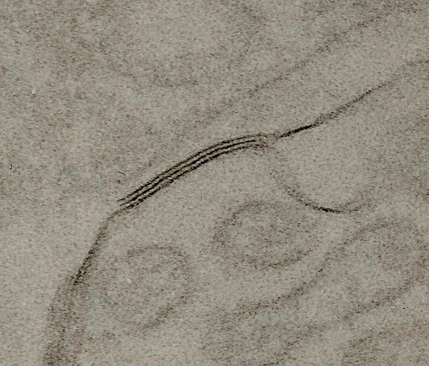
TEM of negatively stained proximal convoluted tubule of Rat kidney tissue at a magnification of ~55,000x and 80 kV with Tight junction. Note that the three dark lines of density correspond to the density of the protein complex, and the light lines in between correspond to the paracellular space.
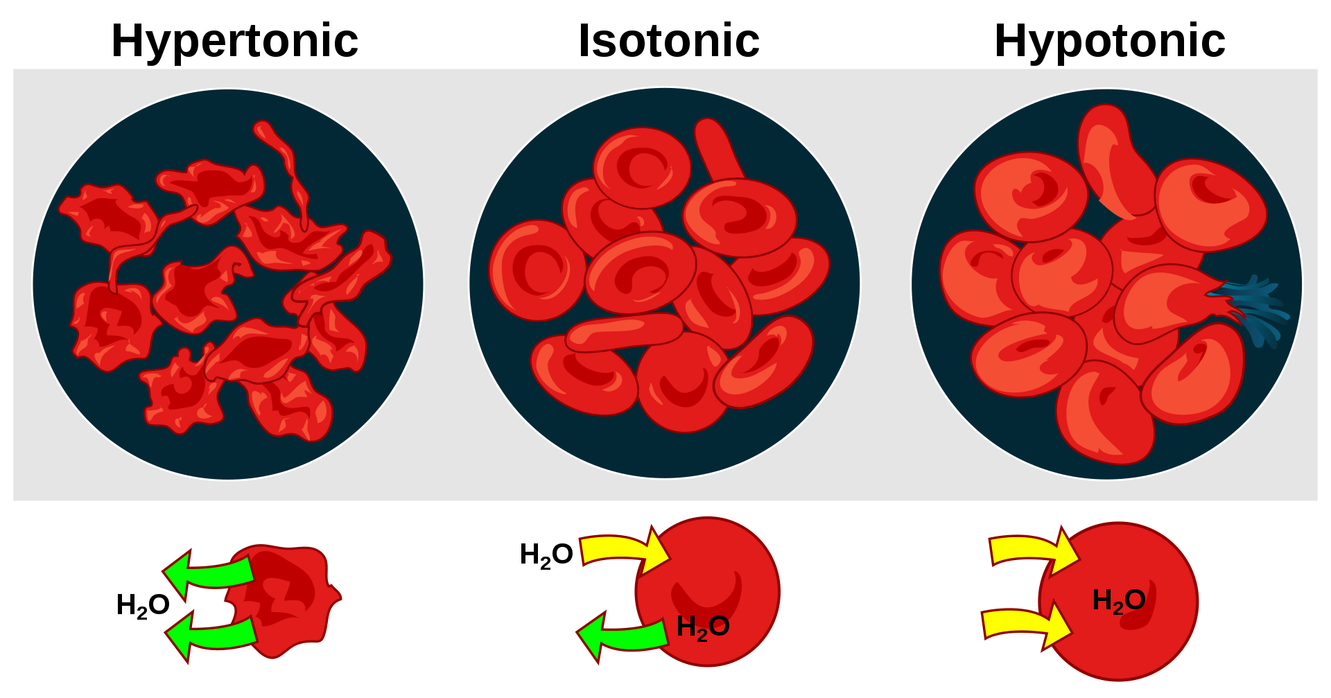
Tonicity is a measure of the effective osmotic pressure gradient; the water potential of two solutions separated by a semipermeable cell membrane. In other words, tonicity is the relative concentration of solutes dissolved in solution which determine the direction and extent of diffusion. It is commonly used when describing the response of cells immersed in an external solution.
Unlike osmotic pressure, tonicity is influenced only by solutes that cannot cross the membrane, as only these exert an effective osmotic pressure. Solutes able to freely cross the membrane do not affect tonicity because they will always equilibrate with equal concentrations on both sides of the membrane without net solvent movement. It is also a factor affecting imbibition.
There are three classifications of tonicity that one solution can have relative to another: hypertonic, hypotonic, and isotonic.(W)
Effect of different solutions on red blood cells.
Osmotic pressure is the hydrostatic pressure produced by a solution in a space divided by a differentially permeable membrane due to a differential in the concentrations of solute.
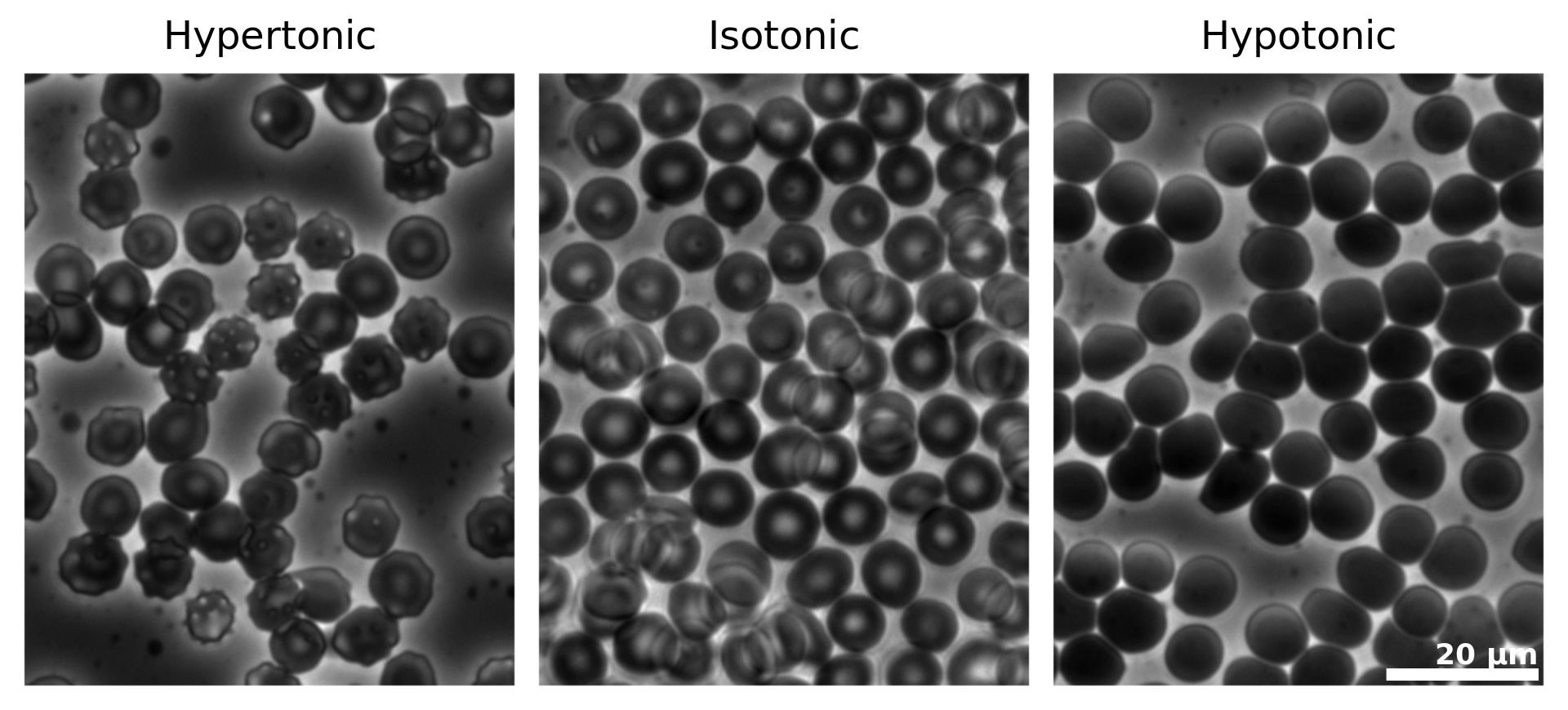
Micrographs of osmotic pressure on red blood cells.
Human erythrocytes (red blood cells) viewed by phase contrast light microscopy. Three conditions are shown: hypertonic conditions (where the erythrocytes contract and appear "spiky"), isotonic conditions (where the erythrocytes appear normal) and hypotonic conditions (where the etrythrocytes expand and become more round).
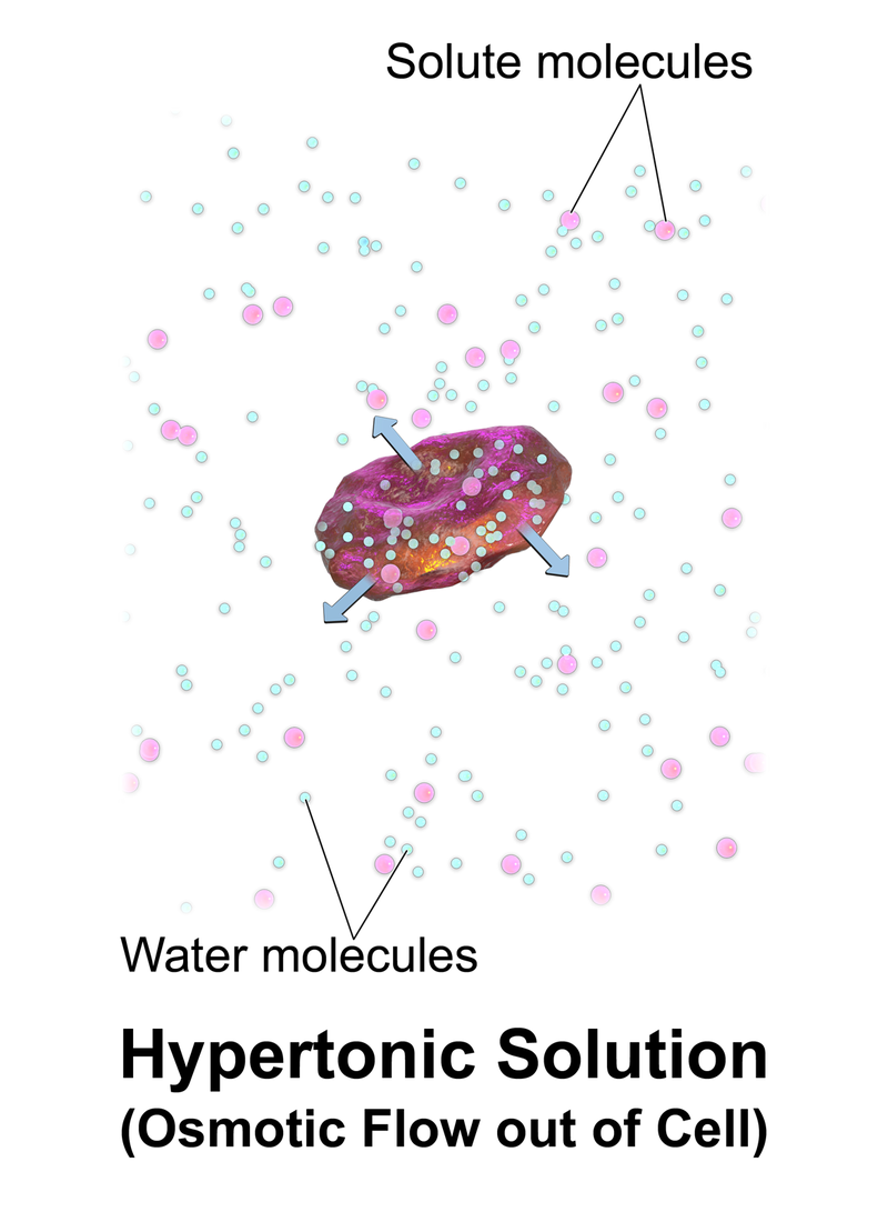
A red blood cell in a hypertonic solution, causing water to move out of the cell.
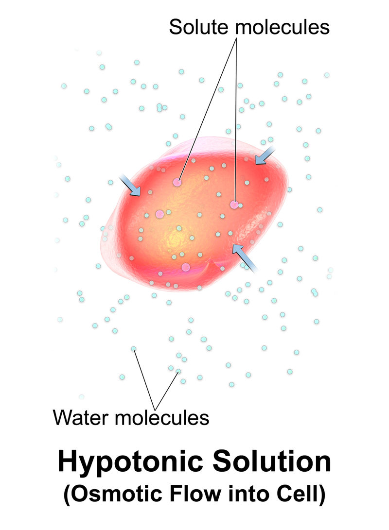
A red blood cell in a hypotonic solution, causing water to move into the cell.
The transcellular pathway of transport is important in the intestinal absorption of drug molecules, the other being the paracellular pathway. The transcellular pathway of transport include transcellular diffusion, active carrier mediated transportation, and transcytosis. The transcelluar diffusion simply involves the movement of solutes based on a diffusion gradient moving from an area of high concentration to an area of low concentration, however, the cell membrane is a hydrophobic environment and will not allow the passive diffusion of charged, hydrophilic, or zwitterion molecules. Active transport involves the use of energy to transport specific substrates across barriers, even against the concentration gradient. Macromolecules can sometimes be transported through transcytosis.
In contrast, paracellular transport is the transfer of substances across an epithelium by passing through an intercellular space between the cells.
1. It differs from transcellular transport, where the substances travel through the cell passing through both the apical membrane and basolateral membrane
2. Renal physiology. Transcellular transport is more likely to involve energy expenditure than paracellular transport.
3. Capillaries of the blood–brain barrier have only transcellular transport, in contrast with normal capillaries, which have both transcellular and paracellular transport. (W)
transformation (genetics)
In molecular biology and genetics,transformation is the genetic alteration of a cell resulting from the direct uptake and incorporation of exogenous genetic material from its surroundings through the cell membrane(s). For transformation to take place, the recipient bacterium must be in a state of competence, which might occur in nature as a time-limited response to environmental conditions such as starvation and cell density, and may also be induced in a laboratory.
Transformation is one of three processes for horizontal gene transfer, in which exogenous genetic material passes from one bacterium to another, the other two being conjugation (transfer of genetic material between two bacterial cells in direct contact) and transduction (injection of foreign DNA by a bacteriophage virus into the host bacterium). In transformation, the genetic material passes through the intervening medium, and uptake is completely dependent on the recipient bacterium.
As of 2014 about 80 species of bacteria were known to be capable of transformation, about evenly divided between Gram-positive and Gram-negative bacteria; the number might be an overestimate since several of the reports are supported by single papers.
"Transformation" may also be used to describe the insertion of new genetic material into nonbacterial cells, including animal and plant cells; however, because "transformation" has a special meaning in relation to animal cells, indicating progression to a cancerous state, the process is usually called "transfection".(W)
In this image, a gene from bacterial cell 1 is moved from bacterial cell 1 to bacterial cell 2. This process of bacterial cell 2 taking up new genetic material is called transformation..
Bacterial Transformation In this diagram, a gene from bacterial cell 1 is moved from bacterial cell 1 to bacterial cell 2. This process of bacterial cell 2 taking up new genetic material is called transformation. Step I: The DNA of a bacterial cell is located in the cytoplasm (1), but also in the plasmid, an independent, circular loop of DNA. The gene to be transferred (4) is located on the plasmid of cell 1 (3), but not on the plasmid of bacterial cell 2 (2). In order to remove the gene from the plasmid of bacterial cell 1, a restriction enzyme (5) is used. The restriction enzyme binds to a specific site on the DNA and “cuts” it, releasing the satisfactory gene. Genes are naturally removed and released into the environment usually after a cell dies and disintegrates. Step II: Bacterial cell 2 takes up the gene. This integration of genetic material from the environment is an evolutionary tool and is common in bacterial cells. Step III: The enzyme DNA ligase (6) adds the gene to the plasmid of bacterial cell 2 by forming chemical bonds between the two segments which join them together. Step IV: The plasmid of bacterial cell 2 now contains the gene from bacterial cell 1 (7). The gene has been transferred from one bacterial cell to another, and transformation is complete. (W)
transgenerational epigenetic inheritance
Transgenerational epigenetic inheritance, (TEI), is the transmission of epigenetic markers from one organism to the next (i.e., parent–child transmission) that affects the traits of offspring without alteration of the primary structure of DNA (i.e. the sequence of nucleotides):168—in other words, epigenetically. The less precise term "epigenetic inheritance" may cover both cell–cell and organism–organism information transfer. Although these two levels of epigenetic inheritance are equivalent in unicellular organisms, they may have distinct mechanisms and evolutionary distinctions in multicellular organisms.
The environment can induce the epigenetic marks (epigenetic tags) for some epigenetically influenced traits, and some marks are heritable, leading some to view epigenetics as a relaxation of biology's rejection of the inheritance of acquired characteristics (Lamarckism).(W)
Translational medicine
Translational medicine (often referred to as translational science, of which it is a form) is defined by the European Society for Translational Medicine (EUSTM) as "an interdisciplinary branch of the biomedical field supported by three main pillars: benchside, bedside, and community". The goal of TM is to combine disciplines, resources, expertise, and techniques within these pillars to promote enhancements in prevention, diagnosis, and therapies. Accordingly, translational medicine is a highly interdisciplinary field, the primary goal of which is to coalesce assets of various natures within the individual pillars in order to improve the global healthcare system significantly. (W)
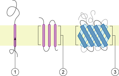
Schematic representation of transmembrane proteins: 1) a single transmembrane α-helix (bitopic membrane protein). 2) a polytopic transmembrane α-helical protein. 3) a polytopic transmembrane β-sheet protein. The membrane is represented in light yellow.
Group I and II transmembrane proteins have opposite final topologies. Group I proteins have the N terminus on the far side and C terminus on the cytosolic side. Group II proteins have the C terminus on the far side and N terminus in the cytosol. However final topology not the only criterion for defining transmembrane protein groups, rather location of topogenic determinants and mechanism of assembly is considered in the classification.
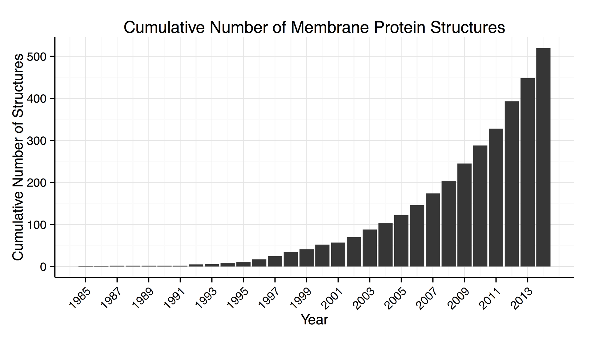
Increase in the number of 3D structures of membrane proteins known.
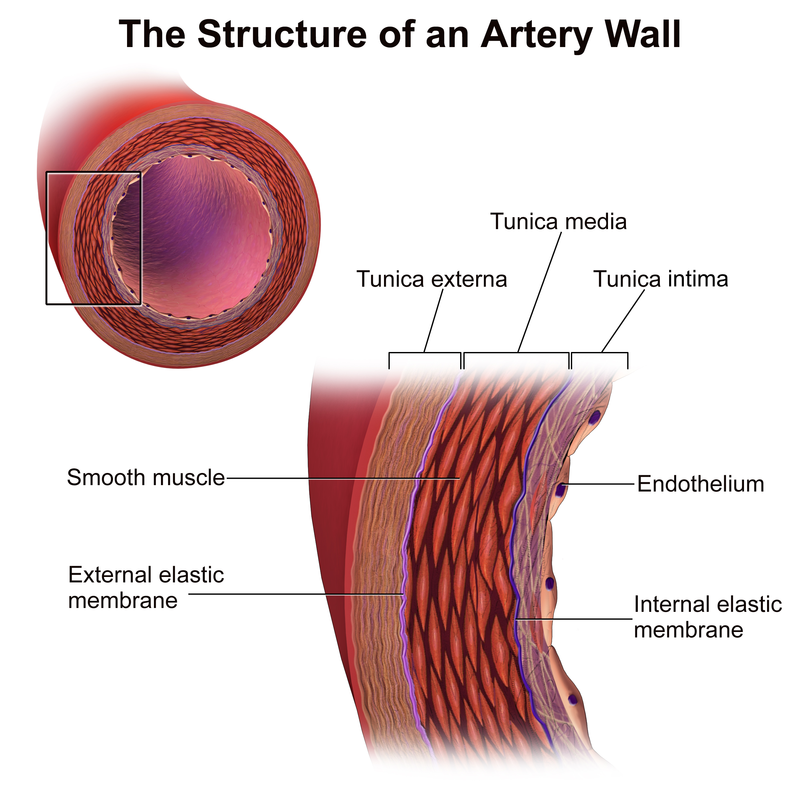
The three layers of a blood vessel are an inner layer (the tunica intima), a middle layer (the tunica media), and an outer layer (the tunica externa).
In dissection, the inner coat (tunica intima) can be separated from the middle (tunica media) by a little maceration, or it may be stripped off in small pieces; but, because of its friability, it cannot be separated as a complete membrane. It is a fine, transparent, colorless structure which is highly elastic, and, after death, is commonly corrugated into longitudinal wrinkles. (W)
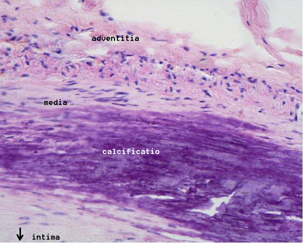
Microphotography of arterial wall with calcified (violet colour) atherosclerotic plaque (haematoxillin and eosin stain).
v
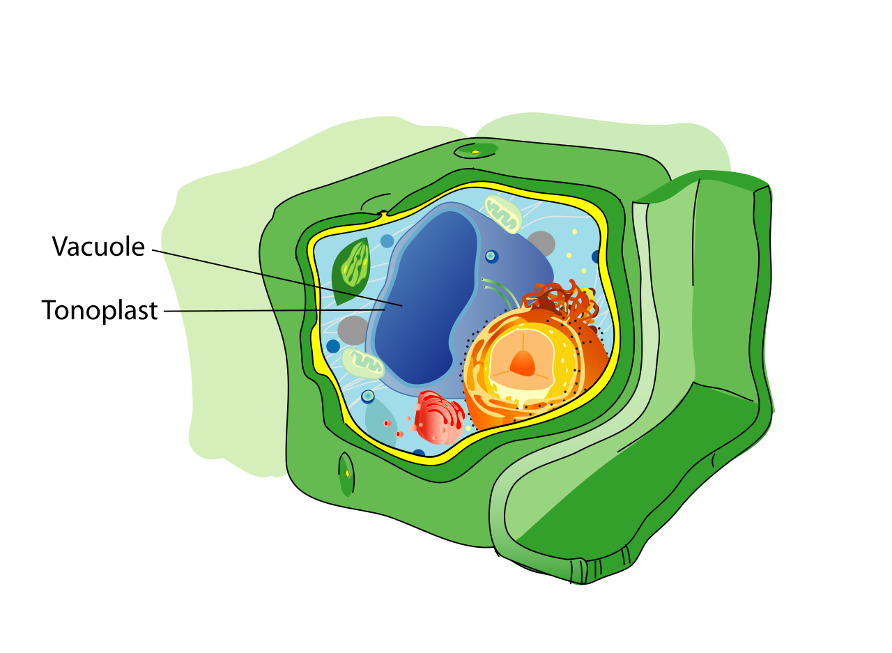
vacuole
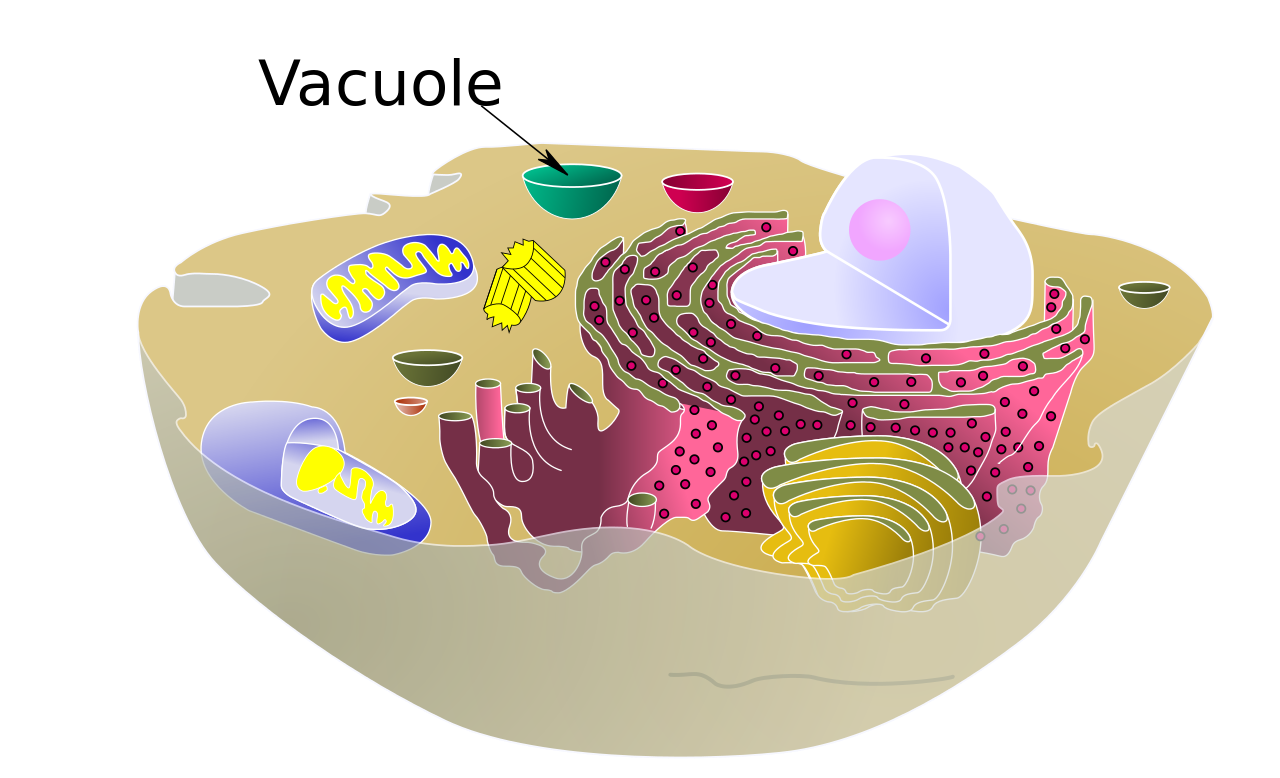
A vacuole is a membrane-bound organelle which is present in plant and fungalcells and some protist,animal and bacterial cells. Vacuoles are essentially enclosed compartments which are filled with water containing inorganic and organic molecules including enzymes in solution,though in certain cases they may contain solids which have been engulfed. Vacuoles are formed by the fusion of multiple membrane vesicles and are effectively just larger forms of these. The organelle has no basic shape or size; its structure varies according to the requirements of the cell. (W)
Plant cell structure.
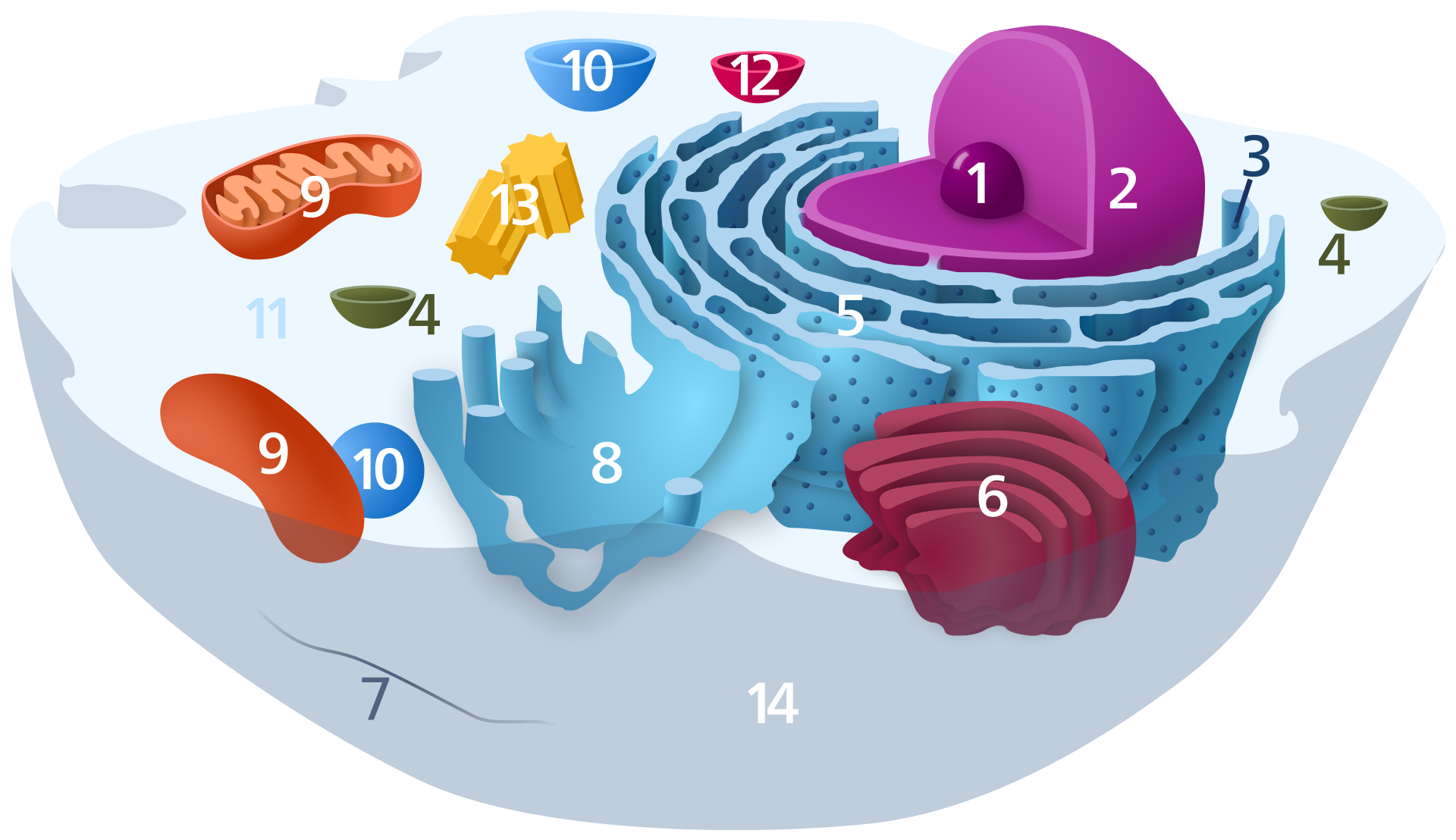
Animal cell structure.
vertebrate
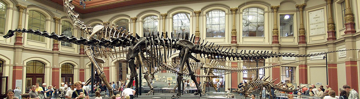
Vertebrates comprise all species of animals within the subphylumVertebrata (chordates with backbones). Vertebrates represent the overwhelming majority of the phylumChordata, with currently about 69,963 species described. Vertebrates include such groups as the following:
The vertebrates traditionally include the hagfish, which do not have proper vertebrae due to their loss in evolution, though their closest living relatives, the lampreys, do. Hagfish do, however, possess a cranium. For this reason, the vertebrate subphylum is sometimes referred to as "Craniata" when discussing morphology. Molecular analysis since 1992 has suggested that hagfish are most closely related to lampreys, and so also are vertebrates in a monophyletic sense. Others consider them a sister group of vertebrates in the common taxon of craniata.
The populations of vertebrates have dropped in the past 50 years. (W)
Fossilized skeleton of Diplodocus carnegii, showing an extreme example of the backbone that characterizes the vertebrates..
vesicle (biology and chemistry)
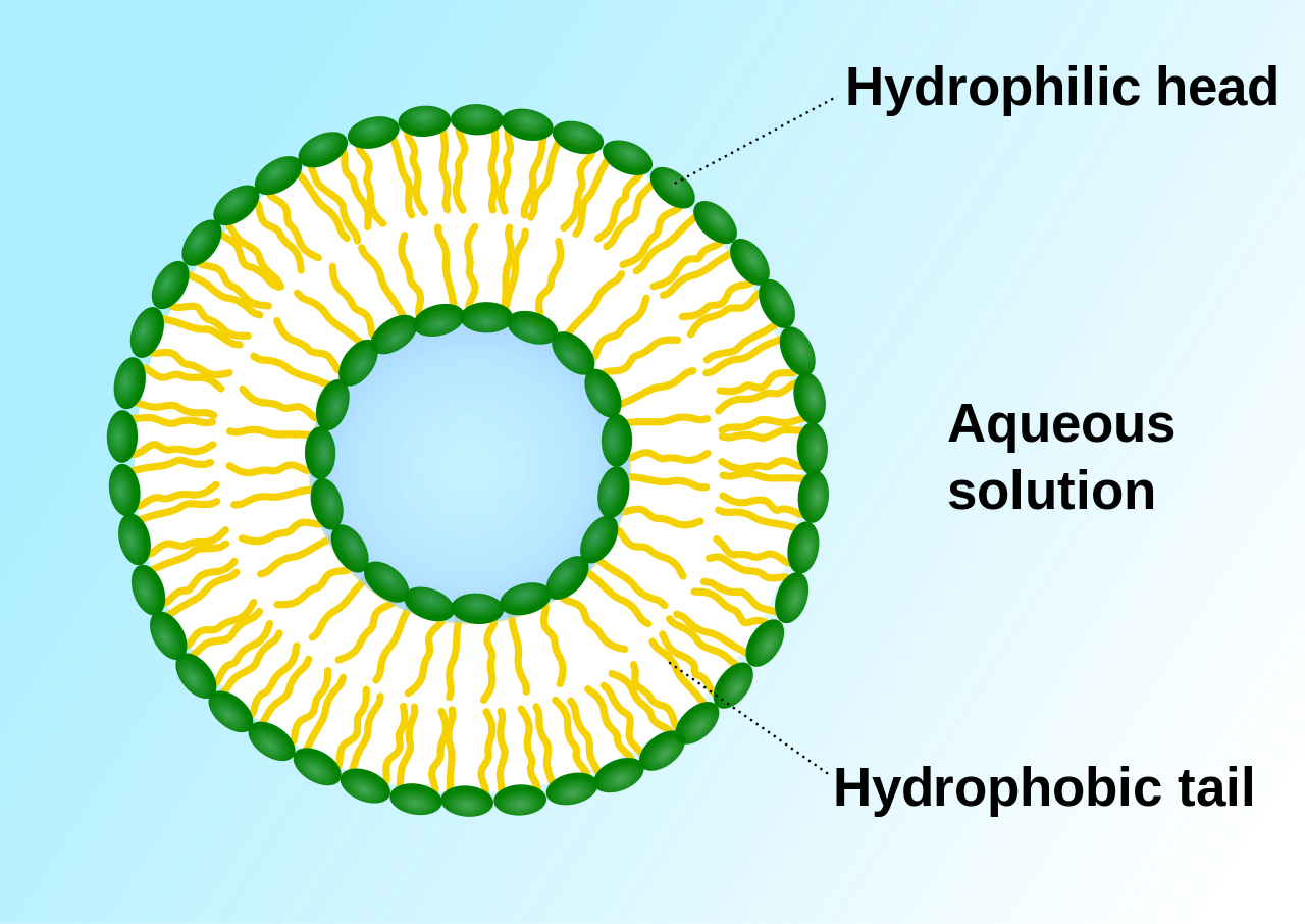
In cell biology, a vesicle is a structure within or outside a cell, consisting of liquid or cytoplasm enclosed by a lipid bilayer. Vesicles form naturally during the processes of secretion (exocytosis) , uptake (endocytosis) and transport of materials within the plasma membrane. Alternatively, they may be prepared artificially, in which case they are called liposomes (not to be confused with lysosomes) . If there is only one phospholipid bilayer, they are called unilamellar liposome vesicles; otherwise they are called multilamellar. The membrane enclosing the vesicle is also a lamellar phase, similar to that of the plasma membrane, and intracellular vesicles can fuse with the plasma membrane to release their contents outside the cell. Vesicles can also fuse with other organelles within the cell. A vesicle released from the cell is known as an extracellular vesicle.(W)
Virophages are small, double-stranded DNA viral phages that require the co-infection of another virus. The co-infecting viruses are typically giant viruses. Virophages rely on the viral replication factory of the co-infecting giant virus for their own replication. One of the characteristics of virophages is that they have a parasitic relationship with the co-infecting virus. Their dependence upon the giant virus for replication often results in the deactivation of the giant viruses. The virophage may improve the recovery and survival of the host organism.
Unlike satellite viruses, virophages have a parasitic effect on their co-infecting virus. Virophages have been observed to render a giant virus inactive and thereby improve the condition of the host organism.
All known virophages are grouped into the family Lavidaviridae (from "large virus dependent or associated" + -viridae).(W)
Lavidaviridae.
Sputnik virophage .
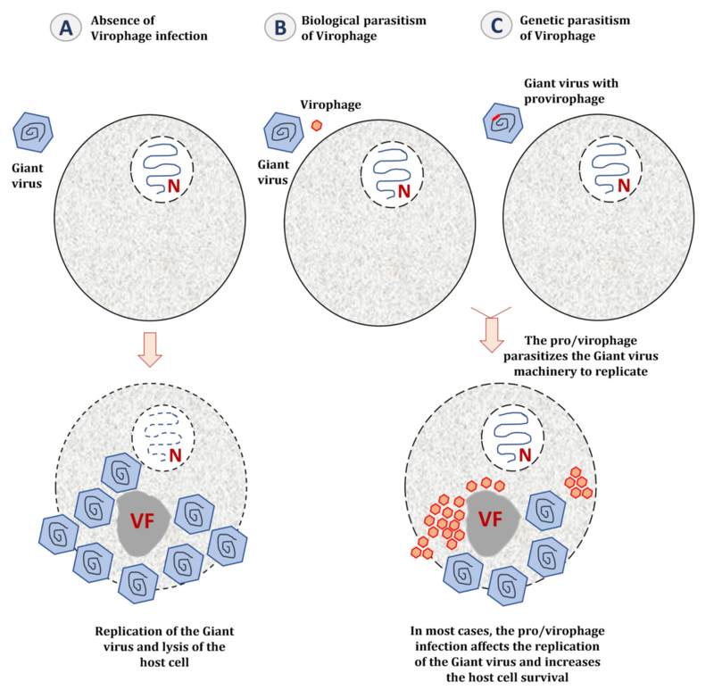
The parasitic lifestyle of virophages (A) When the host cell is only infected by a giant virus, the latter establishes a cytoplasmic virus factory to replicate and generates new virions, and the host cell is most likely lysed at the end of its replication cycle. (B) When the host cell is co-infected with a giant virus and its virophage, the latter parasitizes the giant virus factory. The presence of virophages could seriously impact the infectivity of the giant virus by decreasing its replication efficiency and increasing the survival of the host cell. (C) When the giant virus genome is parasitized by a provirophage, the latter is expressed during the giant virus replication. The virophage is produced from the giant virus factory and inhibits the giant virus replication, thus increasing the host cell survival. VF: Virus factory.
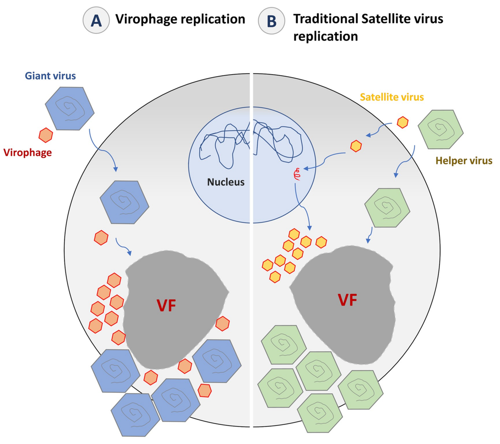
Virophages and satellite virus’ lifestyle (A) The replication of virophages is supposed to occur entirely in the virus factory of its giant virus host, depending of the giant virus expression/replication complex. (B) The concept of satellite virus implicates that the virus initiates the expression and replication of its genome in the nucleus using the host cell machinery and then goes to the cytoplasm. In the cytoplasm, the satellite virus hijacks the morphogenesis machinery of its helper virus to produce its progeny.
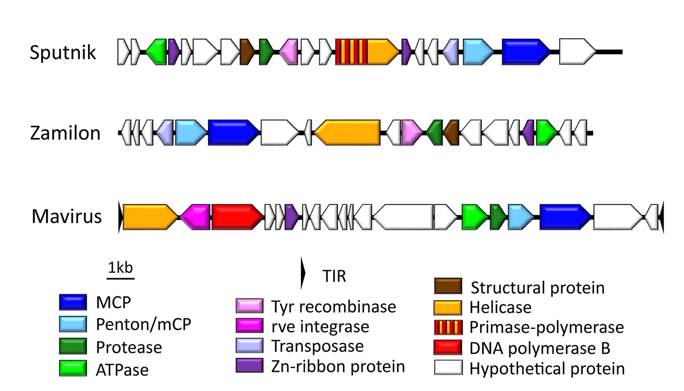
Genome organization of cultured virophages
Genome representation of the virophages Sputnik, Zamilon, and Mavirus. Homologous genes are colored identically..
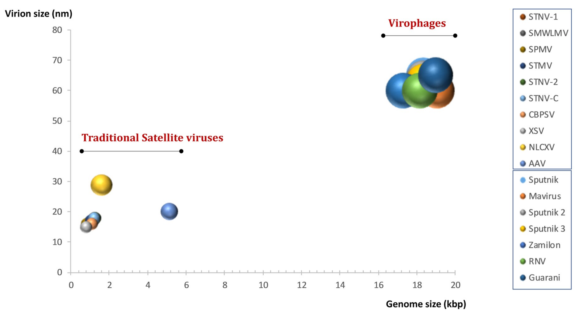
The parasites of the giants are giants Plot comparing the virion and genome sizes for known virophages and some traditional satellite viruses. The ball sizes are proportional to the capsid sizes.
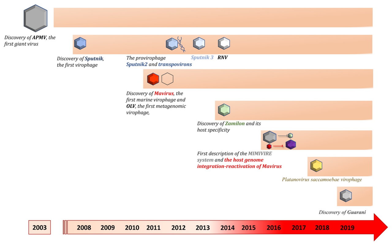
Timeline of virophage discoveries 2003–2019 Timeline showing the chronological order of description of virophages isolated by co-culture and the major discoveries in the virophage field. RNV: Rio Negro Virophage. OLV: Organic Lake Virophage.
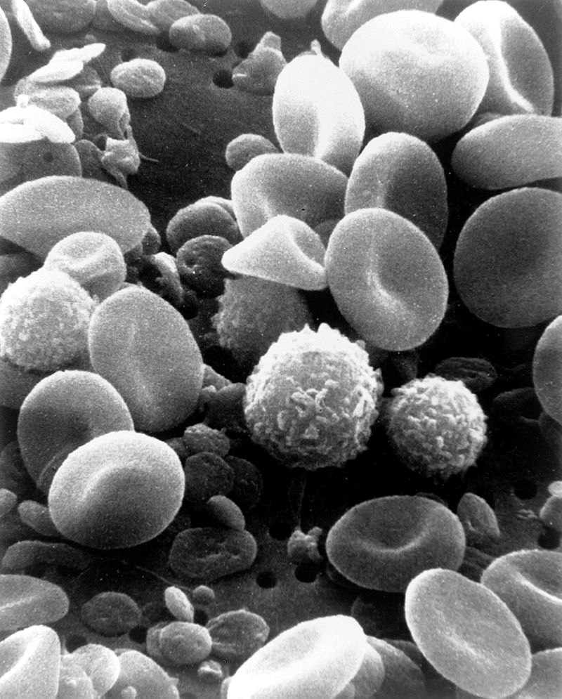
This is a scanning electron microscope image from normal circulating human blood. One can see red blood cells, several white blood cells including lymphocytes, a monocyte, a neutrophil, and many small disc-shaped platelets. Red cells are nonnucleated and contain hemoglobin, an important protein that contains iron and allows the cell to carry oxygen to other parts of the body. They also carry carbon dioxide away from peripheral tissue to the lungs where it can be exhaled. The infection-fighting white blood cells are classified in two main groups: granular and agranular. All blood cells are formed in the bone marrow. There are two types of agranulocytes: lymphocytes, which fight disease by producing antibodies and thus destroying foreign material, and monocytes. Platelets are tiny cells formed in bone marrow and are necessary for blood clotting..
x
X-chromosome inactivation
X-inactivation (also called Lyonization, after English geneticist Mary Lyon) is a process by which one of the copies of the X chromosome is inactivated in therianfemalemammals. The inactive X chromosome is silenced by it being packaged into a transcriptionally inactive structure called heterochromatin. As nearly all female mammals have two X chromosomes, X-inactivation prevents them from having twice as many X chromosome gene products as males, who only possess a single copy of the X chromosome (see dosage compensation).
The choice of which X chromosome will be inactivated is random in placental mammals such as humans, but once an X chromosome is inactivated it will remain inactive throughout the lifetime of the cell and its descendants in the organism. Unlike the random X-inactivation in placental mammals, inactivation in marsupials applies exclusively to the paternally-derived X chromosome. (W)
A 6-year old tortoise shell cat.
The coloration of tortoiseshell and calico cats is a visible manifestation of X-inactivation. The black and orange alleles of a fur coloration gene reside on the X chromosome. For any given patch of fur, the inactivation of an X chromosome that carries one allele results in the fur color of the other, active allele.
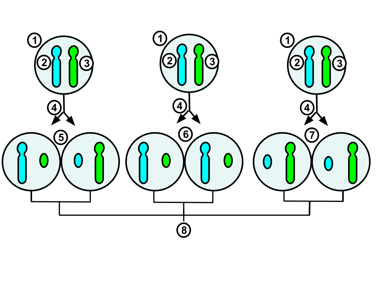
The process and possible outcomes of random X-chromosome inactivation in female human embryonic cells undergoing mitosis. 1.Early stage embryonic cell of a female human 2.Maternal X chromosome 3.Paternal X chromosome 4.Mitosis and random X-chromosome inactivation event 5.Paternal chromosome is randomly inactivated in one daughter cell, maternal chromosome is inactivated in the other 6.Paternal chromosome is randomly inactivated in both daughter cells 7.Maternal chromosome is randomly inactivated in both daughter cells 8.Three possible random combination outcomes.
X-ray crystallography
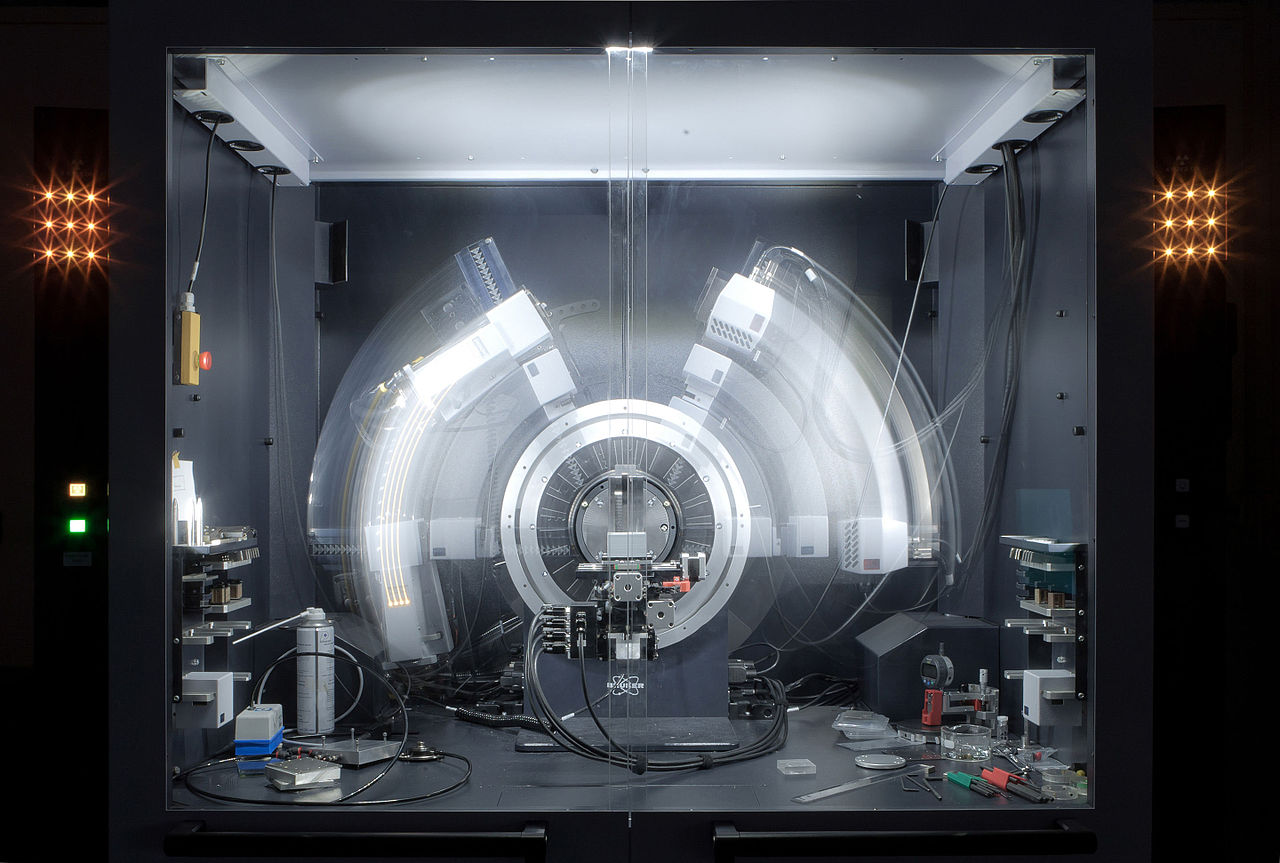
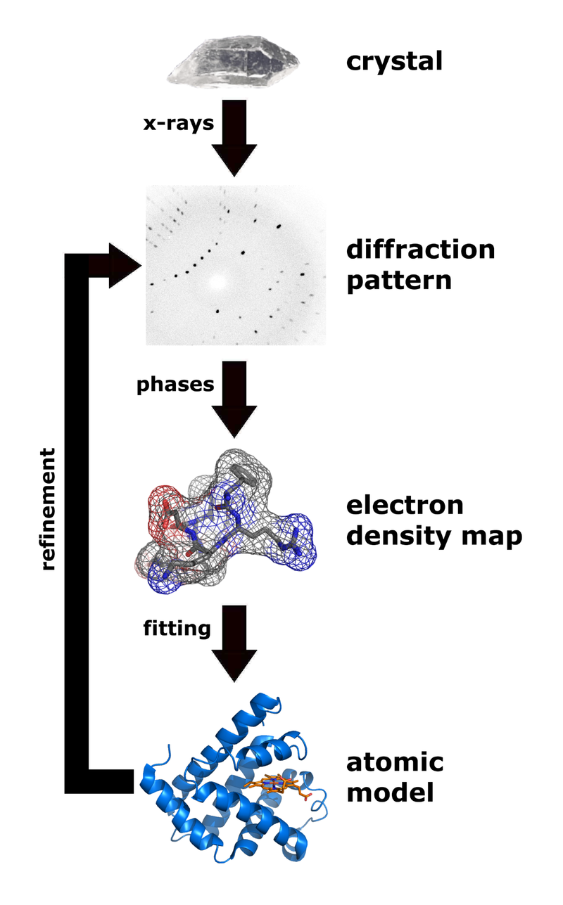
X-ray crystallography (XRC) is the experimental science determining the atomic and molecular structure of a crystal, in which the crystalline structure causes a beam of incident X-rays to diffract into many specific directions. By measuring the angles and intensities of these diffracted beams, a crystallographer can produce a three-dimensional picture of the density of electrons within the crystal. From this electron density, the mean positions of the atoms in the crystal can be determined, as well as their chemical bonds, their crystallographic disorder, and various other information. (W)
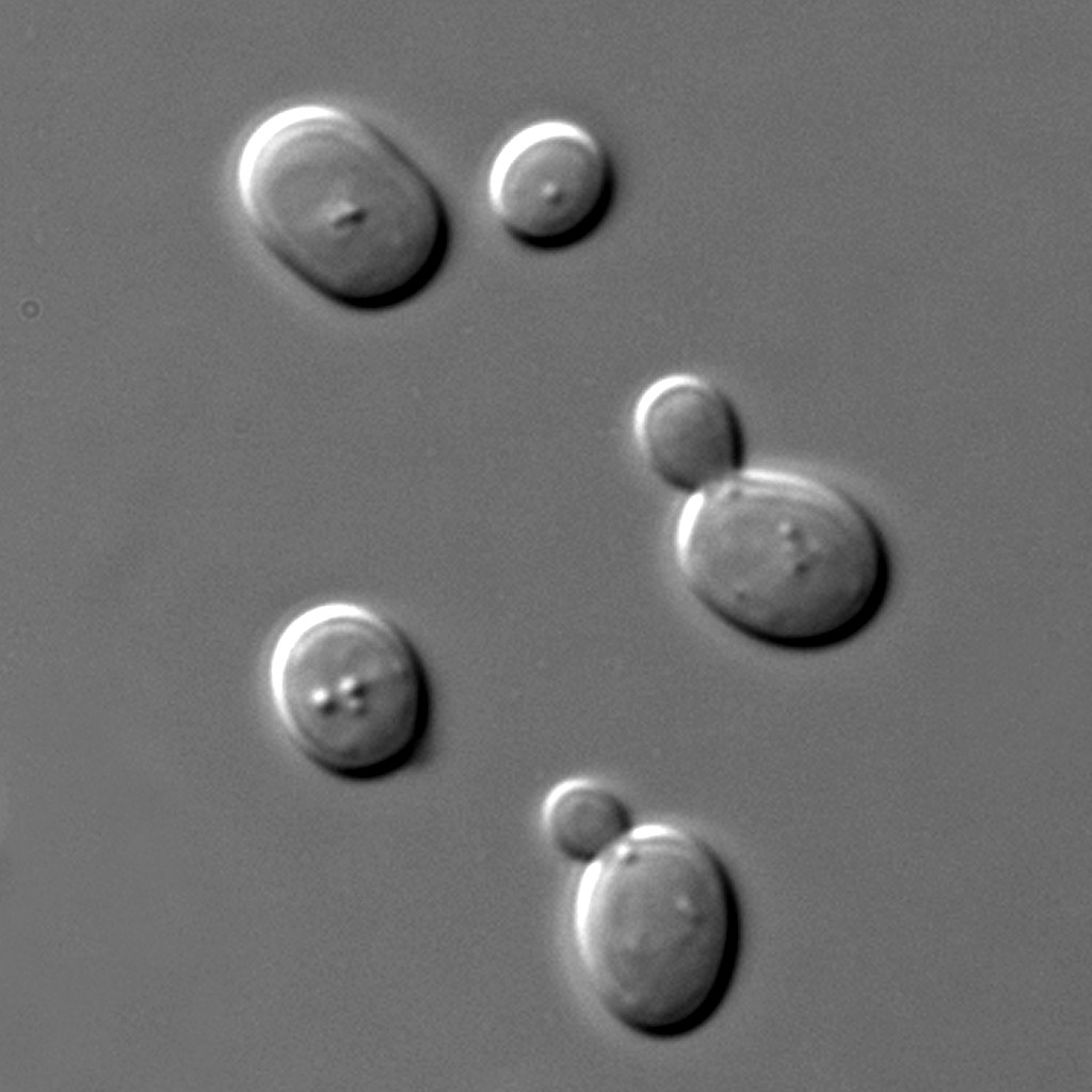
Yeasts are eukaryotic, single-celled microorganisms classified as members of the funguskingdom. The first yeast originated hundreds of millions of years ago, and at least 1,500 species are currently recognized. They are estimated to constitute 1% of all described fungal species. (W)
Saccharomyces cerevisiae cells in DIC microscopy. Imaging was performed with the Olympus BX61 microscope and a UPlanSApo 100× NA 1.40 oil immersion objective (Olympus). Pictures were acquired at room temperature in synthetic complete medium with a camera (SPOT; Diagnostic Instruments, Inc.) using MetaMorph software (MDS Analytical Technologies).
Cross-sectional 2D diagram of a yeast cell.
z
zygote
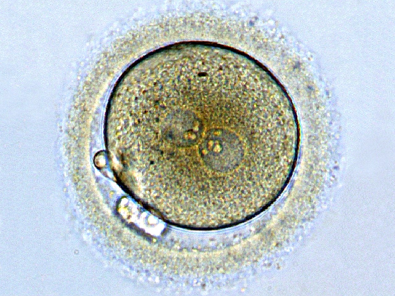
A zygote (from Greek ζυγωτός zygōtos "joined" or "yoked", from ζυγοῦν zygoun "to join" or "to yoke") is a eukaryoticcell formed by a fertilization event between two gametes. The zygote's genome is a combination of the DNA in each gamete, and contains all of the genetic information necessary to form a new individual. In multicellular organisms, the zygote is the earliest developmental stage. In single-celled organisms, the zygote can divide asexually by mitosis to produce identical offspring.
German zoologists Oscar and Richard Hertwig made some of the first discoveries on animal zygote formation in the late 19th century. (W)
Zygote: egg cell after fertilization with a sperm. The male and female pronuclei are converging, but the genetic material is not yet united.


.jpg)
.png)


.png)